Figure 2. Microphotographs of the
immunolabeling of newborn pig ciliary epithelium (CE)-derived
cells after in vitro differentiation on poly-D-Lysine, laminin
coated coverslips in the presence of 1% serum and 10 ng/ml basic
fibroblast growth factor (bFGF) and epidermal growth factor
(EGF). The images were acquired using an epifluorescent
microscope. A, B: Cells labeled for recoverin
are clustered together (arrows). C:
4',6-diamidino-2-phenylindole (DAPI) nuclear staining
corresponding to A and B. D, E:
Cells double-labeled for recoverin (red, D) and
rhodopsin (green, D and E) are depicted by
arrows. The focus is set to show rhodopsin-positive cell
processes. Strong recoverin staining in the cytoplasm masks the
nuclear DAPI staining, which is shown separately in F.
Cells positive for protein kinase α (PKCα; green in G
and H, arrows) did not co-label with recoverin (red in G
and H and another example in the inset in G,
arrowheads). The focus is set to show the processes of
PKCα-labeled cells in G and H, and the
recoverin-labeled processes in the inset in G.
Corresponding DAPI nuclear stain is shown in I. J-K:
A calbindin immunopositive cell is depicted by the arrow. The
focus is set to show the processes of the labeled cell.
Corresponding DAPI nuclear stain is shown in L. M,
N: Recoverin-positive cells (arrows in M and N)
that had retained pigmented granules (arrow in the bright-field
image in O). P, Q: RPE-65 immunopositive
cells (arrows) and the corresponding nuclear DAPI staining in R.
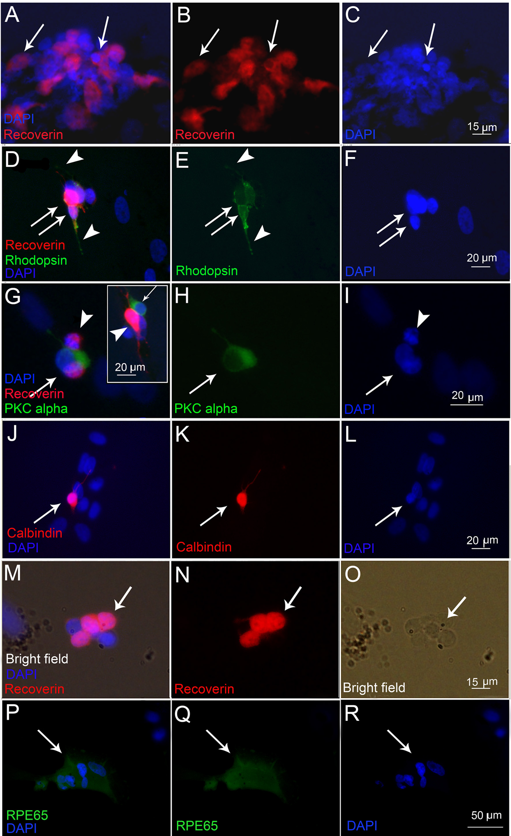
 Figure 2
of Guduric-Fuchs, Mol Vis 2011;
17:2580-2595.
Figure 2
of Guduric-Fuchs, Mol Vis 2011;
17:2580-2595.