Figure 4. Family 3: two affected
brothers, affected paternal aunt. A: At one and one-half
year of age, the proband developed left acute pupillary block
glaucoma. Complete crystalline lens dislocation into the
anterior chamber of the left eye and a large corneal diameter
(14 mm horizontally) can be seen. B: The proband's
six-month-old brother was tentatively scheduled for primary
congenital glaucoma surgery by his physician. Megalocornea is
evident (14 mm horizontal diameter without breaks or scarring).
The child also had bilateral spherophakia (not shown). C:
The paternal aunt of the proband had been diagnosed with
glaucoma at ten years of age but never had surgery. At 20 years
old, bilateral symmetric megalocornea (14 mm horizontal diameter
without breaks or scarring) is evident. Both crystalline lenses
were posteriorly dislocated (not shown). She had high
intraocular pressure, angle synechiae, and glaucomatous optic
nerve damage in her right eye (not shown).
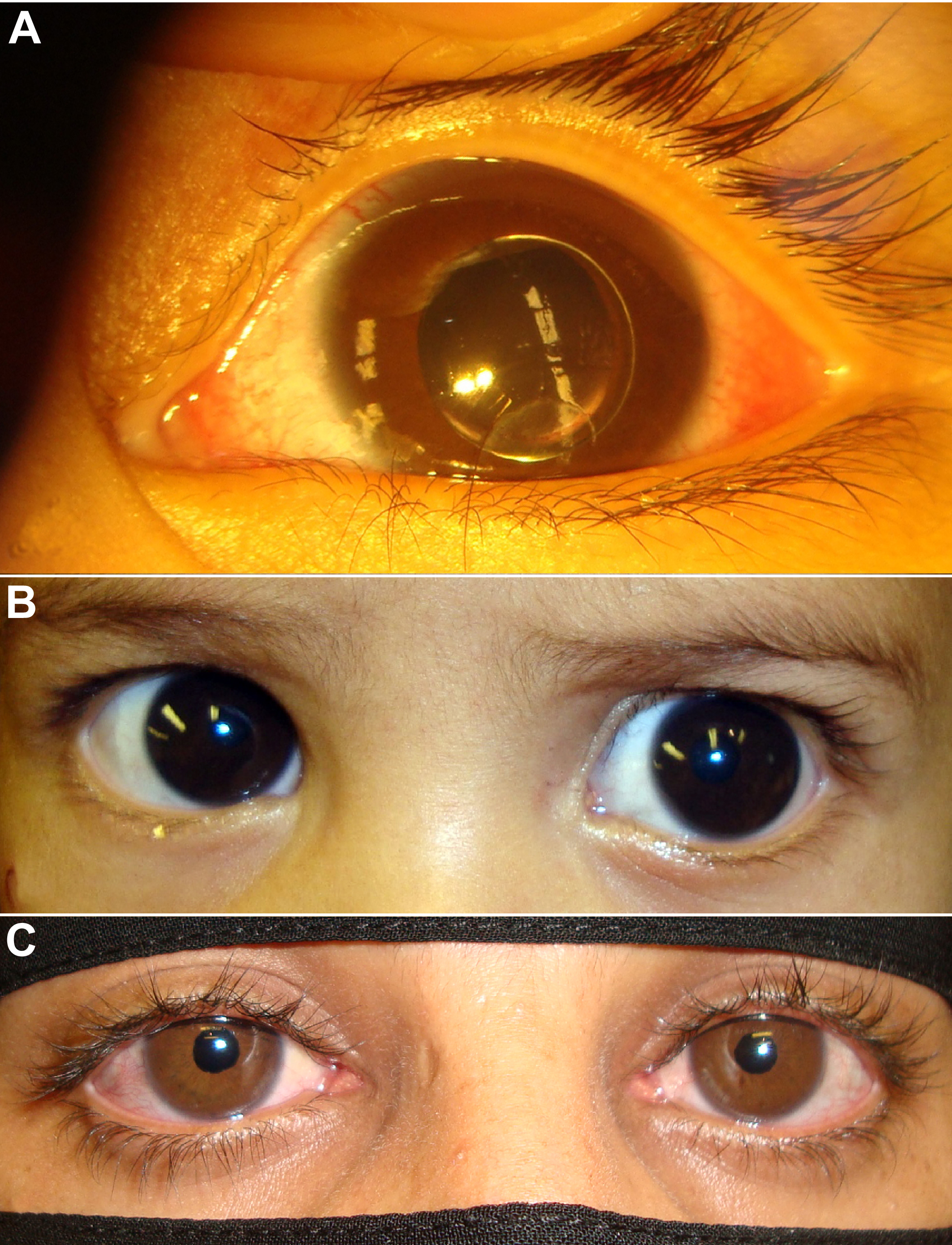
 Figure 4
of Khan, Mol Vis 2011; 17:2570-2579.
Figure 4
of Khan, Mol Vis 2011; 17:2570-2579.