Figure 2. Fundus photographs of the right eye of the 38-year-old choroideremia-affected proband of family 1 show symmetric profound
chorioretinal atrophy with preservation of the central macula. A: The fundus shows areas of retinal pigment epithelium (RPE) disruption, severe chorioretinal atrophy, loss of choriocapillaris,
and bare sclera. B: Fluorescein angiography of the same affected male patient shows extensive chorioretinal atrophy with preservation of an
island of RPE at the macular area.
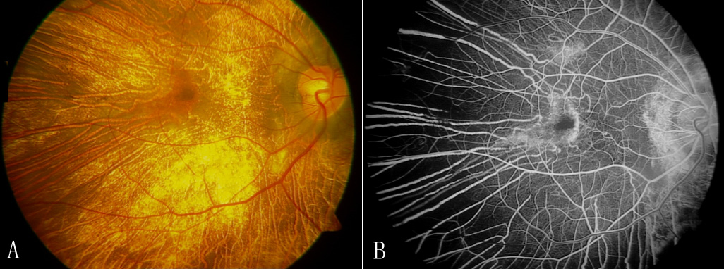
 Figure 2 of
Lin, Mol Vis 2011; 17:2564-2569.
Figure 2 of
Lin, Mol Vis 2011; 17:2564-2569.