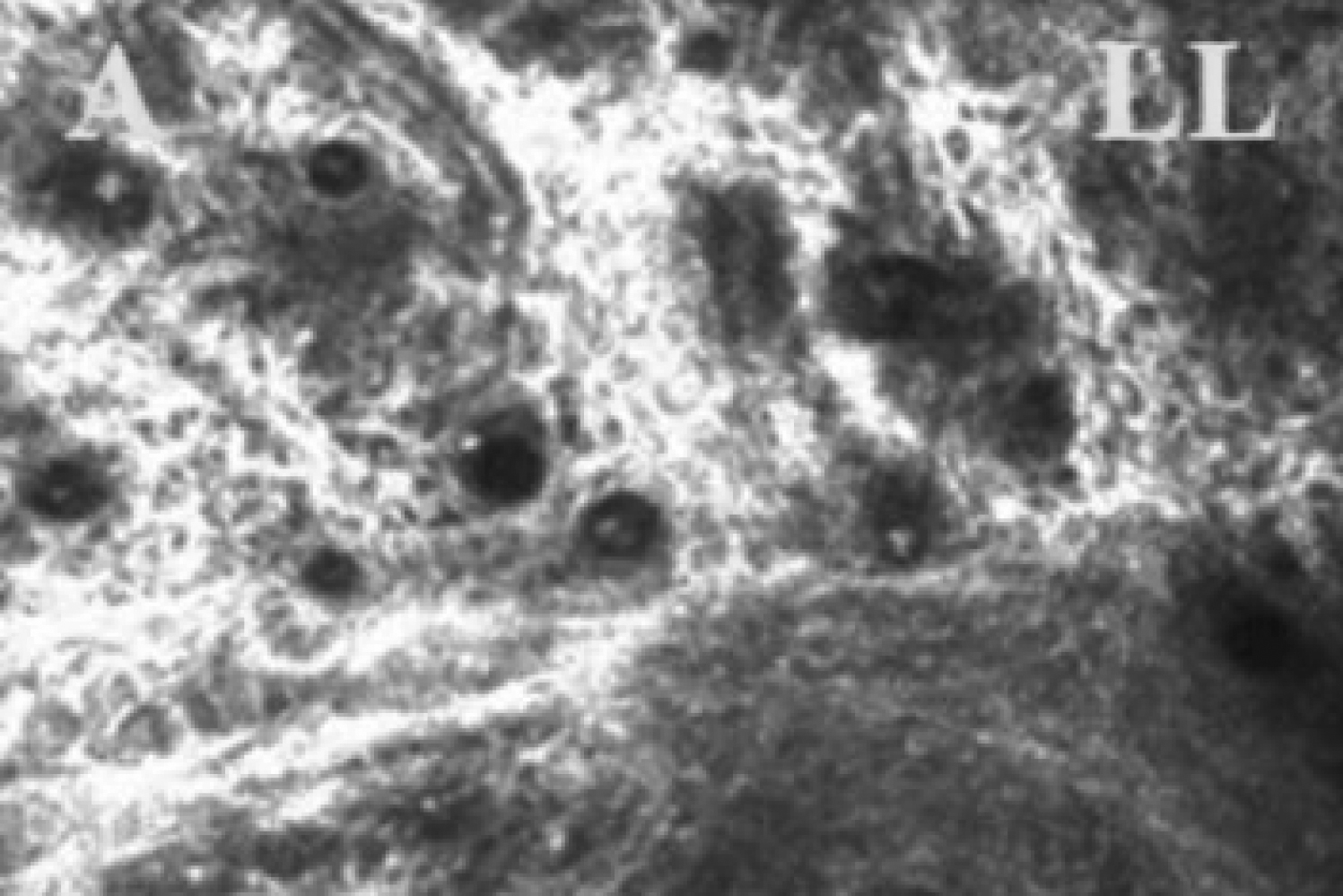
Figure 4. Meibomian gland of DE/cGVHD
patient images observed by in vivo laser confocal microscopy.
DE/cGVHD group, 55-year-old male (Case 9;
Table 1). The
images observed after 17 months on the onset of DE related to
cGVHD. Note the excessive fibrosis around the atrophic glands
and the mild infiltration of inflammatory cells in the dry eye
patients with cGVHD. LL=Lower, Left.
 Figure 4
of Ban, Mol Vis 2011; 17:2533-2543.
Figure 4
of Ban, Mol Vis 2011; 17:2533-2543.