Figure 3. Alignment of protein sequences of human, mouse and zebrafish VSX2/Vsx2/vsx2. The homeodomain sequence is highlighted in green
and the CVC motif in blue. The positions of the mutations identified in Patients 1 (P1) and 2 (P2) are marked in red.
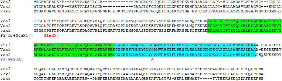
 Figure 3 of
Reis, Mol Vis 2011; 17:2527-2532.
Figure 3 of
Reis, Mol Vis 2011; 17:2527-2532.