Figure 7. Proliferating cell nuclear
antigen (PCNA) analysis in each group at 28 days post-op. A-D:
Immunohistochemistry for PCNA: A: Group A, B:
Group B, C: Group C, D: Group D. E:
Quantitation of the number of PCNA positive cells in the
subconjunctival space at post-op 28 days. *p<0.01 for treated
(rapamycin) versus control (Group B and D; n=4). Original
magnification: 200×.
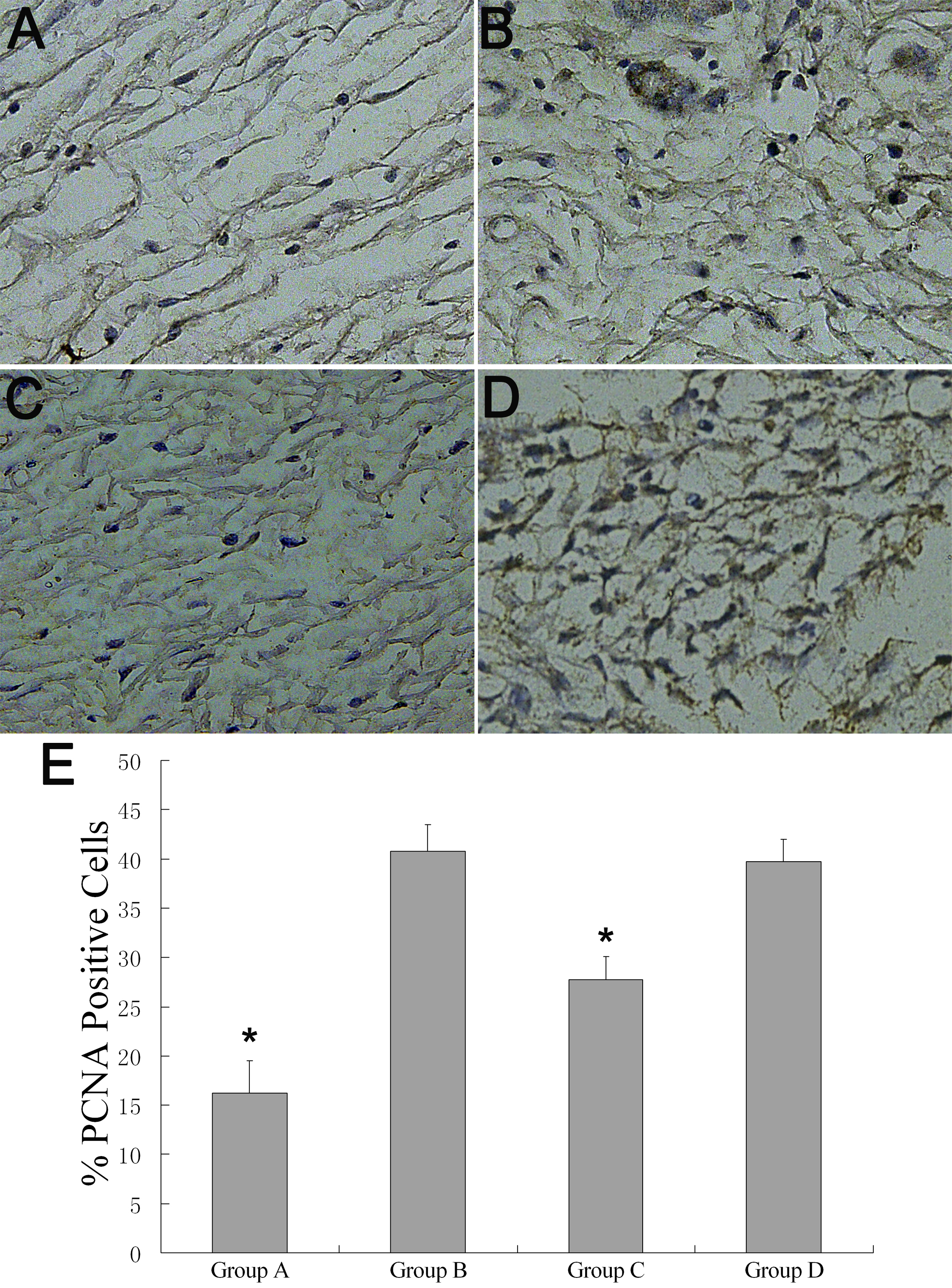
 Figure 7
of Yan, Mol Vis 2011; 17:2495-2506.
Figure 7
of Yan, Mol Vis 2011; 17:2495-2506.