Figure 6. Histopathologic features of
the surgical area in each group at 28 days post-op. A-H
represent pathological changes in the surgical area of Group A,
Group B, Group C, and Group D at 28 days post-op, respectively.
Conjunctiva Epithelium (CE) in each group are in good condition,
conjunctiva flap and scleral (S) space are wide in A and
B, and the Tenon’s Capsule (TC) has few fibroblasts and
new collagen tissue. In C-H conjunctiva flap and
scleral space narrow or even disappear, and a large amount of
fibroblasts hyperplasia and new collagen tissue deposition can
be observed in the Tenon’s Capsule (TC). *represents
sustained-release drug film area. H-E staining, original
magnification: A 40×; C 40×; E 100×; G
100×. Masson trichrome staining, original magnification: B
40×; D 100×; F 100×; H 40×.
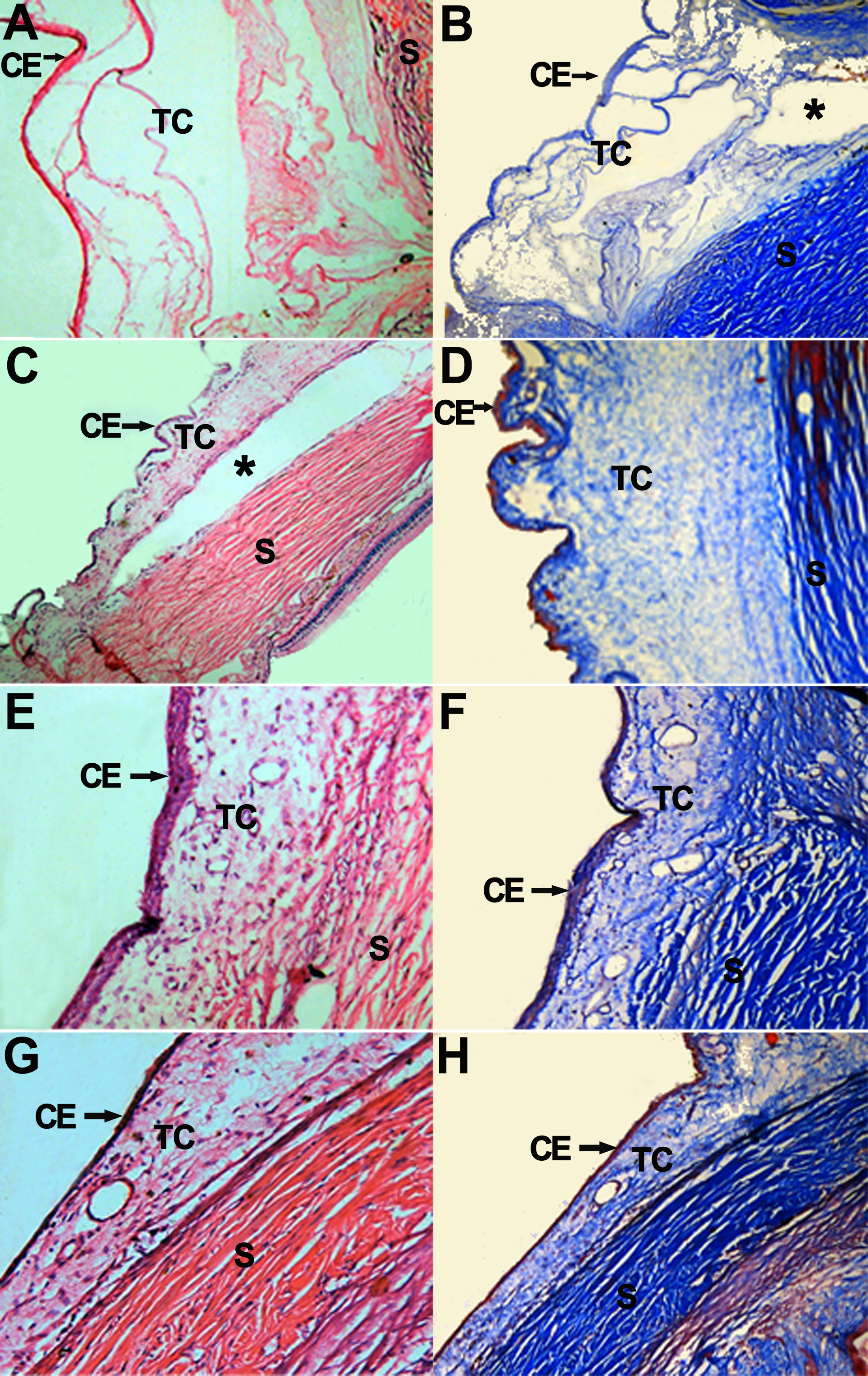
 Figure 6
of Yan, Mol Vis 2011; 17:2495-2506.
Figure 6
of Yan, Mol Vis 2011; 17:2495-2506.