Figure 5. Histopathologic features of
the surgical area in different groups. A, E: At
day 7 and day 14 post-op in Group A, respectively, showing
intact conjunctiva epithelium (CE), only mild inflammation
infiltrate in subconjunctiva scleral filtration area, wide
conjunctiva and scleral space (S), and small amounts of
fibroblasts in Tenon’s Capsule (TC). B, E: Group
B and Group D showing that conjunctival epithelium (CE) is
intact, infiltration of inflammation around the subconjunctival
scleral filtration area, conjunctiva flap and sclera (S) space
narrowing, and dense fibroblasts hyperplasia in the Tenon’s
Capsule (TC) at 7 days post-op. *represents sustained-release
drug film area in B. C: Showing a moderate
number of fibroblasts hyperplasia and a certain subconjunctiva
space at day 7 post-op In Group C, however, F, shows
dense fibroblasts hyperplasia in the Tenon’s Capsule (TC) and
subconjunctival space almost disappear at day 14 post-op. (H-E
staining, original magnification: A 40×; B 200×;
C 200×; D 200×; E 100×; F 100×).
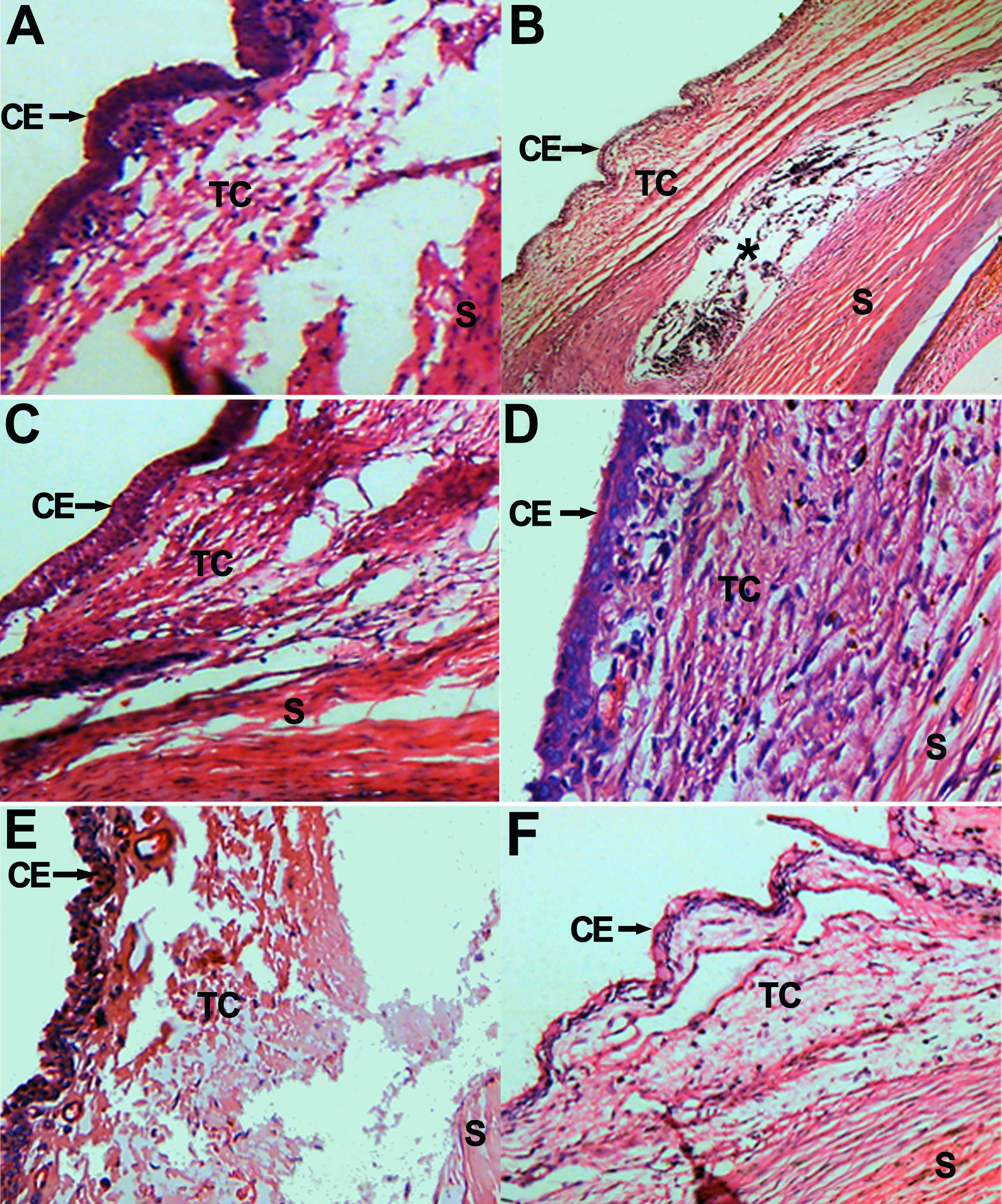
 Figure 5
of Yan, Mol Vis 2011; 17:2495-2506.
Figure 5
of Yan, Mol Vis 2011; 17:2495-2506.