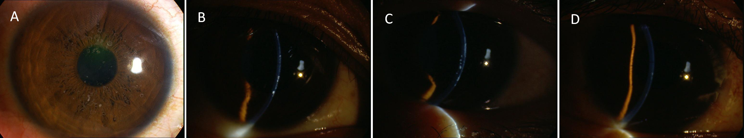
Figure 2. Clinical photographs of patients
with atypical CDGG1.
A: A slit lamp photograph of the proband's
cornea (
Figure 1, V2)
showing
multiple round and dot-like grayish white opacities in the
central stroma.
B: A slit lamp photograph of the proband's
cornea showing the opacities in the epithelium and anterior stroma.
C:
A
slit lamp photograph of the cornea in patient IV6 (
Figure 1, IV6)
showing the opacities in the epithelium and total stroma.
D: A
slit lamp photograph of the cornea in patient IV20 (
Figure 1,
IV20) showing opacities only in the epithelium.
 Figure 2 of Zhu, Mol Vis 2011; 17:225-230.