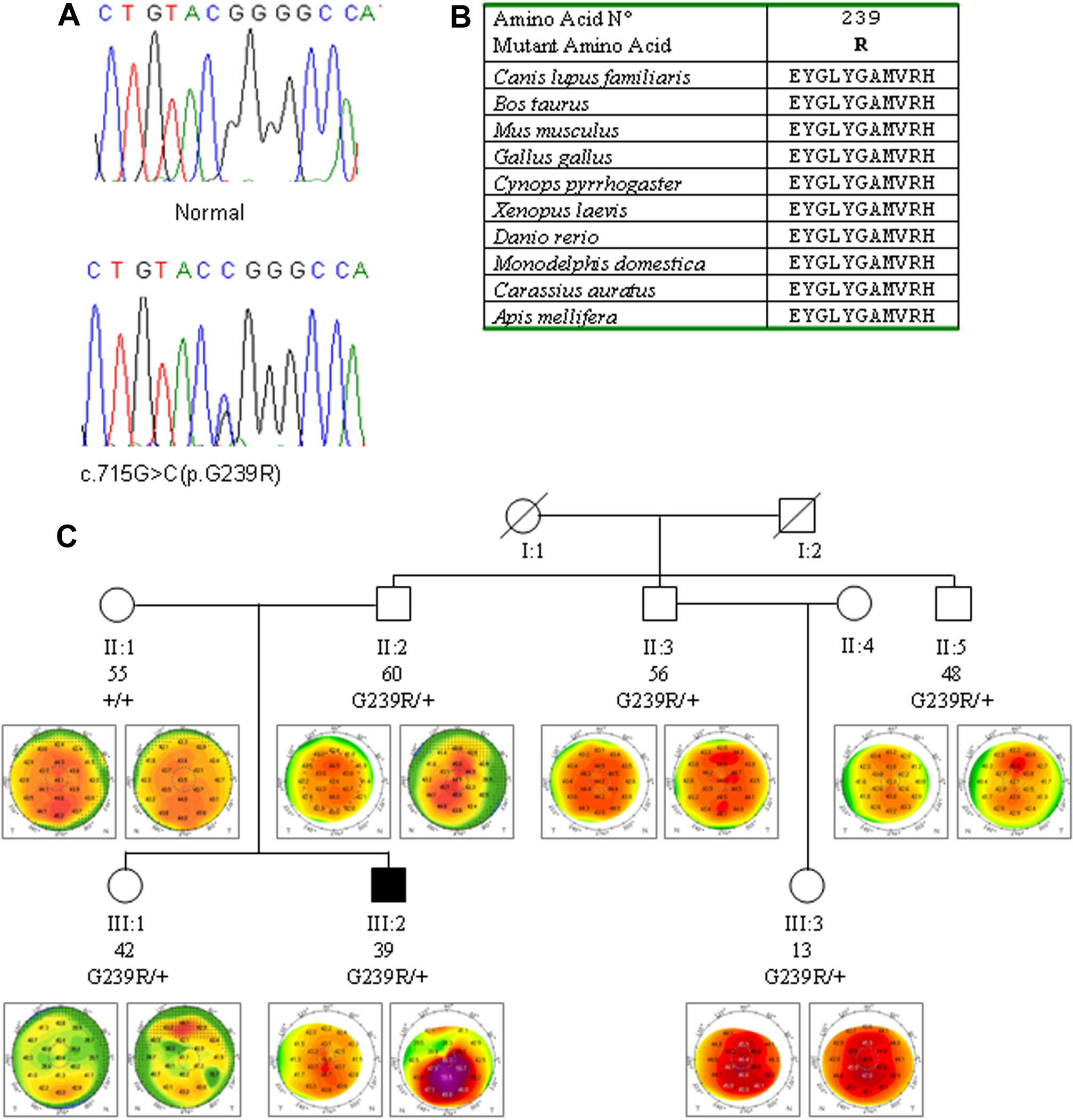
Figure 1. Analysis of the c.715G>C
(p.G239R) sequence variant in
VSX1 exon 4.
A:
DNA sequence electropherogram of the c.715G>C (p.G239R)
sequence variant in
VSX1 exon 4 (
NM_014588).
B: multiple sequence alignment of the amino acid
sequences of VSX1 in different species. Alignments were
performed using the program
Clustal (provided in
the public domain by European Bioinformatics Institute, European
Molecular Biology Laboratory, Heidelberg, Germany).
C:
segregation of p.G239R in family K264. Each individual was
reported by age (in years), genotype and videokeratographs.
Filled symbols refer to keratoconus individual, whereas open
symbols are individuals without clinical keratoconus.
 Figure 1
of De Bonis, Mol Vis 2011; 17:2482-2494.
Figure 1
of De Bonis, Mol Vis 2011; 17:2482-2494.