Figure 5. Neuronal re-innervation of
the corneal epithelium occurs rapidly after injury. Transverse
sections (A-E, H-O) or whole-mount
preparations (F-J) were labeled with Draq5 (red)
or antibodies to SV2 (green) and substance P (red; N and
O). The inset in B is a high magnification view
of SV2-positive neuronal ending in the basal layers of the
corneal epithelium. Panel E is a twofold enlargement of
the boxed-out (yellow rectangle) area in D. Panel F
is a projection of confocal Z-stack of images taken from the
surface of the epithelium down to 80 µm of depth. Panel G
is a projection of confocal Z-stack of images from 80 µm down to
140 µm of depth from the surface of the cornea. Panel H
is a 3D shadow reconstruction to demonstrate the arrangement of
SV2-positive nerve terminals re-innervating regenerated
epithelium. Panels I and J are 20° and 90°
rotations of confocal Z-stack reconstructions to demonstrate the
re-innervation of the trough of regenerated epithelium within
the corneal incision. Arrows indicate SV2/substance P-positive
axons in the stroma. The calibration bar (50 µm) in panel O
applies to panels A-D, K, L, and
M-O. The bar in H applies to all F-H
panels, the bar in I applies to I alone, the bar
in J applies to J alone. Abbreviations: epi –
epithelium, str – stroma, endo – endothelium.
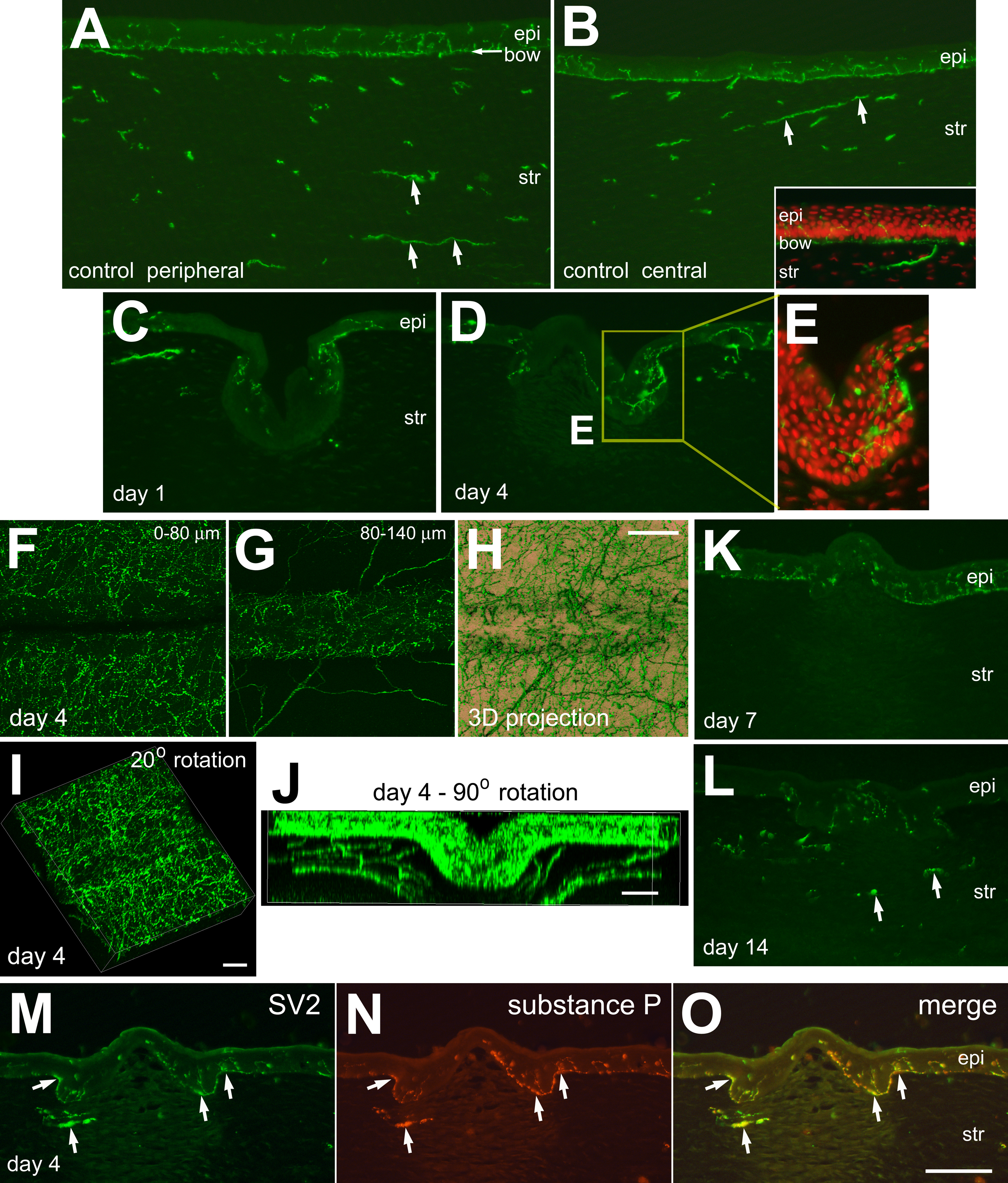
 Figure 5
of Ritchey, Mol Vis 2011; 17:2440-2454.
Figure 5
of Ritchey, Mol Vis 2011; 17:2440-2454.