Figure 4. CD45+ monocytes accumulate
in the stroma following corneal injury. Transverse section of
undamaged (A and B) and injured (C-L)
corneas were labeled with Draq5 (red nuclei in A-I;
blue nuclei J and L) and antibodies to CD45
(green) and BrdU (red in J and L). Corneas were
harvest at 1 (C, G, and J), 4 (D,
E, and L), 7 (F), and 14 (H and I)
days after injury. Panel G is a 2.5 fold enlargement of
the area (yellow box) in C indicating the amoeboid
morphology of CD45+ monocytes in the site of injury in the
stroma. Panel K is a 1.5 fold enlargement of the area
(yellow box) in J providing a single-channel images and
a merged overlay of labeling for CD45, BrdU and Draq5 in a
single monocyte. Arrows indicate cells labeled for CD45 and
Draq5, arrowheads indicate BrdU-labeled cells (J and L),
and small double-arrows indicate a single cell labeled for CD45,
BrdU and Draq5 (J and K). The calibration bar (50
µm) in panel A applies to A alone, the bar in I
applies to B-I, and the bar in L applies
to J and L. Abbreviations: epi – epithelium, str
– stroma, endo – endothelium.
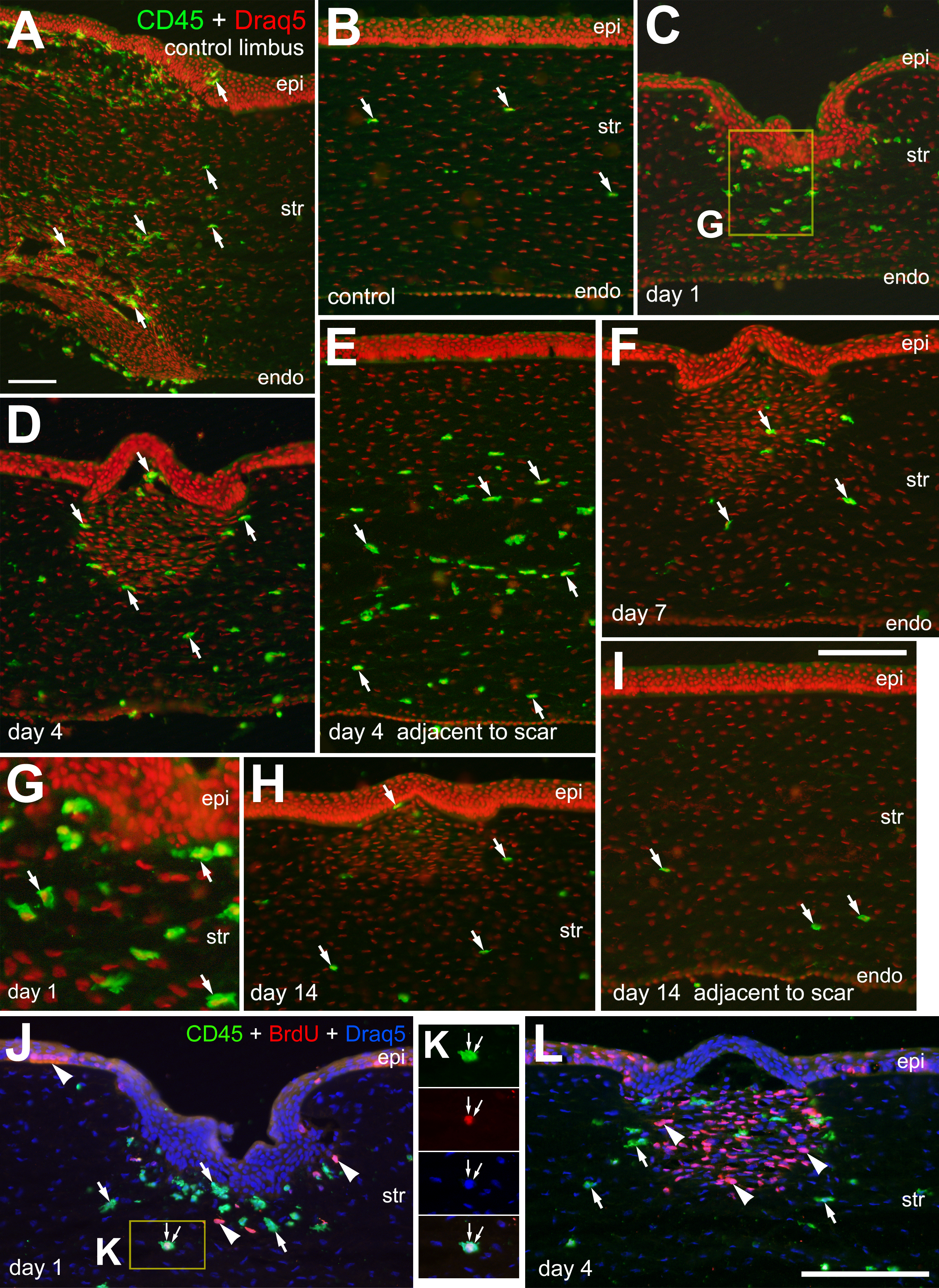
 Figure 4
of Ritchey, Mol Vis 2011; 17:2440-2454.
Figure 4
of Ritchey, Mol Vis 2011; 17:2440-2454.