Figure 1. Corneal injury stimulates
the proliferation of stromal cells. Transverse sections of the
cornea were labeled for BrdU (green) and Draq5 (red nuclei).
Uninjured corneas (A-C) and injured corneas were
harvested (and BrdU injected) at day 1 (BrdU injected 4 h prior;
D), day 4 (BrdU injected at days 1, 2, and 3; E),
day 7 (BrdU injected at days 4 and 5; F), and day 14
(BrdU injected at days 7 and 8; G). Arrows indicate
BrdU-labeled nuclei. H: Plot indicating the mean (±SD;
n=3 for each time point) number of BrdU-labeled cells within
20,000 µm2 within the stroma within the region of the
incision. The calibration bar (50 µm) in G applies to A-G.
I: Plots indicating the mean (±SD; n≥5 for each time
point) indicate the mean number of stromal nuclei per 20,000 µm2
in control, undamaged corneas (red), treated stromal region
adjacent to the incision site (green), and treated stromal
region within the incision site (blue). ANOVA was performed to
determine significance of difference within the data set and a
Bonferroni post-hoc comparison was performed to determine
significance between and within experimental groups. There was
no significant difference (p>0.05) over time among treatment
groups, with the exception of between 1 and 4 days after injury
within the site of incision (p=0.002; I). Abbreviations:
epi – epithelium, str – stroma, endo – endothelium.
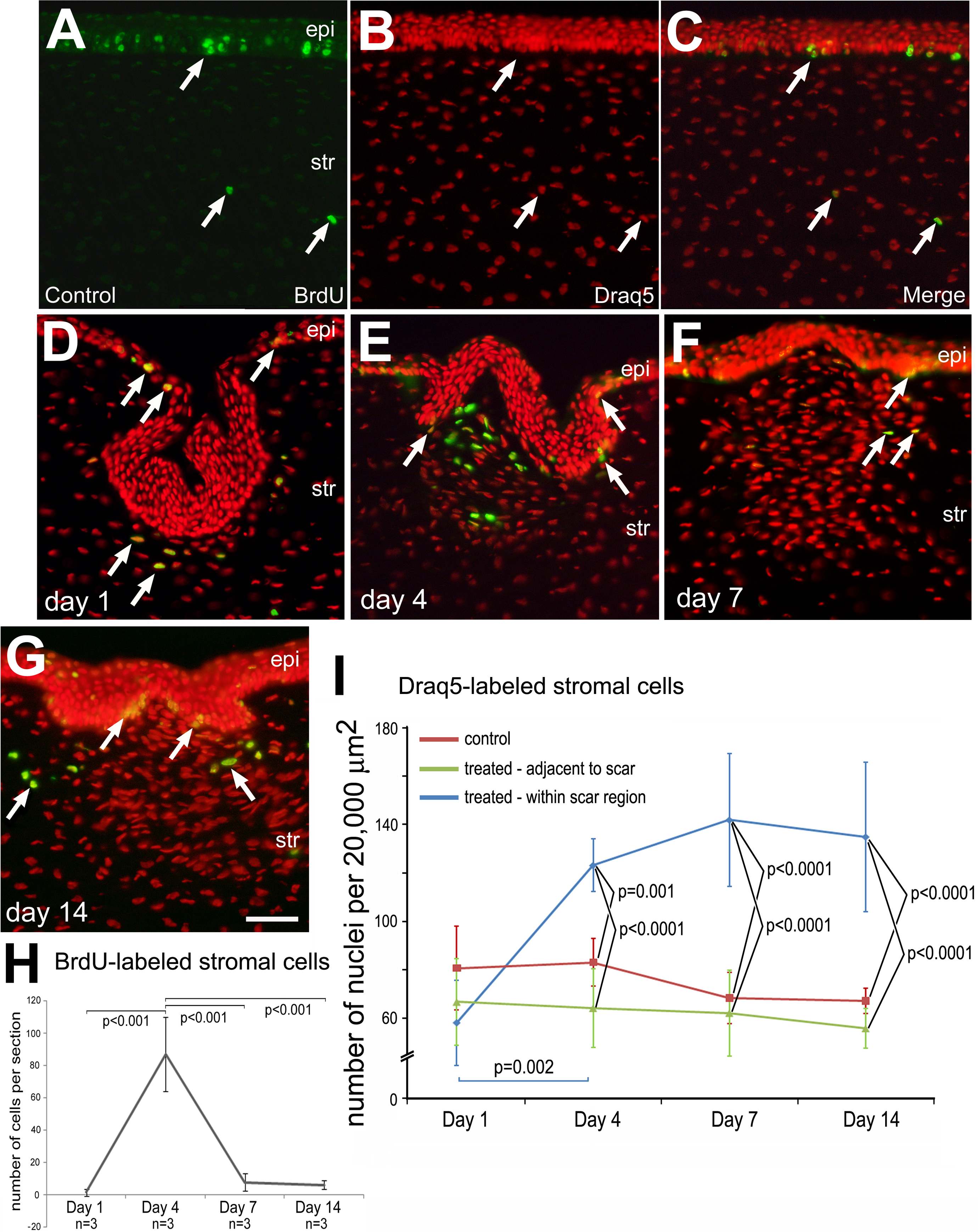
 Figure 1
of Ritchey, Mol Vis 2011; 17:2440-2454.
Figure 1
of Ritchey, Mol Vis 2011; 17:2440-2454.