Figure 2. Photographs of the proband. A and B display the slit lamp photographs of both of the proband’s eyes after the pupils were dilated, showing ectopia lentis. In
the right eye (A), the lens is dislocated nasally. In the left eye (B), the lens is dislocated superonasally. C shows arachnodactyly of the proband.
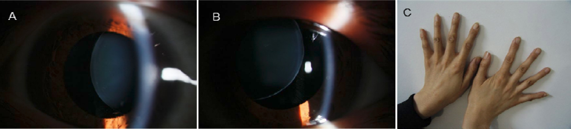
 Figure 2 of
Meng, Mol Vis 2011; 17:2421-2427.
Figure 2 of
Meng, Mol Vis 2011; 17:2421-2427.