Figure 3. Change of expression of PEDF and VEGF detected by western blot in corneal neovascularization. Samples were harvested at day
5 after S-CorNV induction and proteins of equivalent to one fifth cornea were loaded and detected by western blot. Please
note that due to differential levels of two factors in samples, the exposing time of blotted membrane against X-films varied,
namely about 45 s for PEDF, 2 min for GAPDH, and about 1 h for VEGF. Shown was one representative of three experiments that
gave similar conclusions.
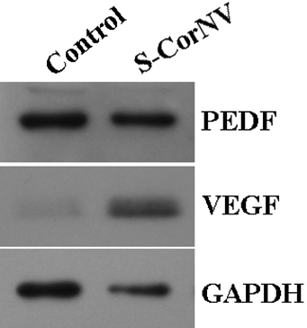
 Figure 3 of
Jia, Mol Vis 2011; 17:2386-2399.
Figure 3 of
Jia, Mol Vis 2011; 17:2386-2399.