Figure 1. Gross and histology presentations of corneas of corneal neovascularization models. A: Front views and side views of corneas with corneal neovascularization under slit lamp. B: Hematoxylin-eosin staining of corneas at limbus area and junction area, the latter of which refers to the area of the suture
stitch in the S-corneal neovascularization model and the margin of direct chemical burn. The red scale bar represent 50 μm.
Please note the difference of manifestations in these two models, especially in terms of transparency of the corneas.
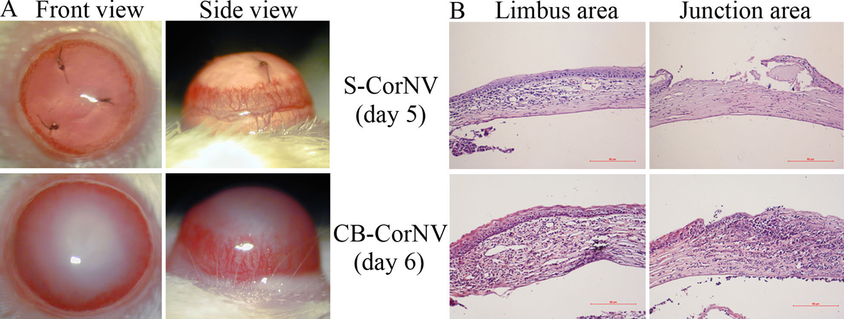
 Figure 1 of
Jia, Mol Vis 2011; 17:2386-2399.
Figure 1 of
Jia, Mol Vis 2011; 17:2386-2399.