Figure 3. Western blots of α subunits from whole LG homogenates. Both α1 and α2 were significantly increased in LG from rabbits with
IAD (p<0.05). β-Actin was used as loading controls. Data are representative images of at least 3 different animals each.
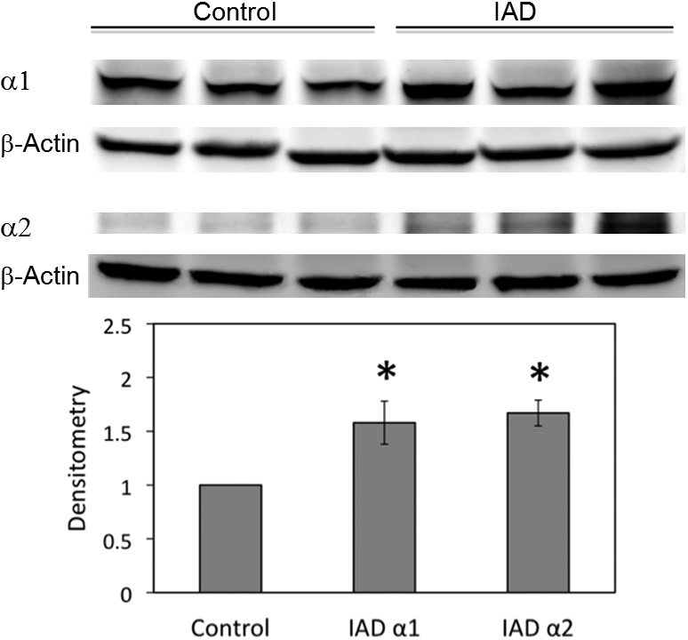
 Figure 3 of
Ding, Mol Vis 2011; 17:2368-2379.
Figure 3 of
Ding, Mol Vis 2011; 17:2368-2379.