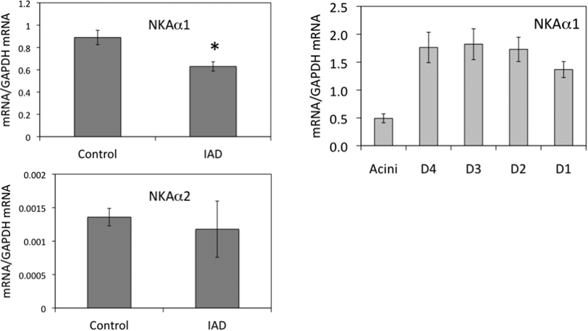
Figure 1. Real-time RT–PCR of α1 and
α2 from whole LG (left panels) and lacrimal epithelial cells
collected by LCM (right panel). mRNA levels of α1 from whole LG
of IAD rabbits were significantly lower than those of control
animals (p<0.05), whereas no significant difference was
detected between control and IAD animals for α2 (p>0.05). In
epithelial cells collected by LCM, mRNA of α1 was the least
abundant in acini, whereas its level was significantly higher in
all duct segments. Compared to control animals [
13],
mRNA level of α1 was significantly lower in acini and every duct
segment except in intralobar duct in IAD animals (p<0.05).
However, we were unable to detect any NKAα2 in epithelial cells
collected by LCM due to its low level. D4: intralobular duct.
D3: interlobular duct. D2: intralobar duct. D1: interlobar duct.
Data were presented as mean±SEM.
 Figure 1
of Ding, Mol Vis 2011; 17:2368-2379.
Figure 1
of Ding, Mol Vis 2011; 17:2368-2379.