Figure 3. Representative images of
histopathologic analysis of four corneal sections – three after
penetrating keratoplasty and one after deep anterior lamellar
keratoplasty. A: Section of the cornea after deep
anterior lamellar keratoplasty. Male patient (F1; 37 years old).
Green birefringence is visible with a polarizing filter
(arrowheads). Stromal deposition of amyloid substance in
anterior corneal part distorts the architecture of the corneal
lamellae. The absence of Bowman’s layer and thinning of the
epithelium are noticeable. LCDI/R124C mutation. B:
Section of the cornea after penetrating keratoplasty. Congo red
stain. Female patient (F2; 45 years). Deposits throughout the
corneal stroma stain positive with Congo red. Note the deep,
posterior corneal location of the deposit (arrowhead). LCD
variant/H626 mutation. C: Section of the cornea after
penetrating keratoplasty. PAS stain. Female patient (F4; 53
years). Note the absence of Bowman’s layer and the distorted
epithelium in correspondence of the granular deposits
(arrowheads). There are several granular deposits throughout the
corneal stroma (arrows). GCDI/R555W mutation. D: Section
of the cornea after penetrating keratoplasty. PAS stain. Female
patient (F4; 53 years). Masson trichrome stain. Section of the
cornea showing the absence of Bowman’s layer and the absence of
the epithelium in correspondence with the Masson trichrome –
positive granular deposits (arrows). GCDI/R555W mutation. E:
Section of the cornea after penetrating keratoplasty. Masson
trichrome stain. Female patient (F9; 44 years). Note that the
granular deposits are placed under the thinner epithelium, thus
taking the place of the former Bowman’s layer. GCDII/R124H
mutation. F: Section of the cornea after penetrating
keratoplasty. Congo red stain. Female patient (F9; 44 years).
Note the Congo red positive deposits in the anterior corneal
stroma. GCDII/R124H mutation.
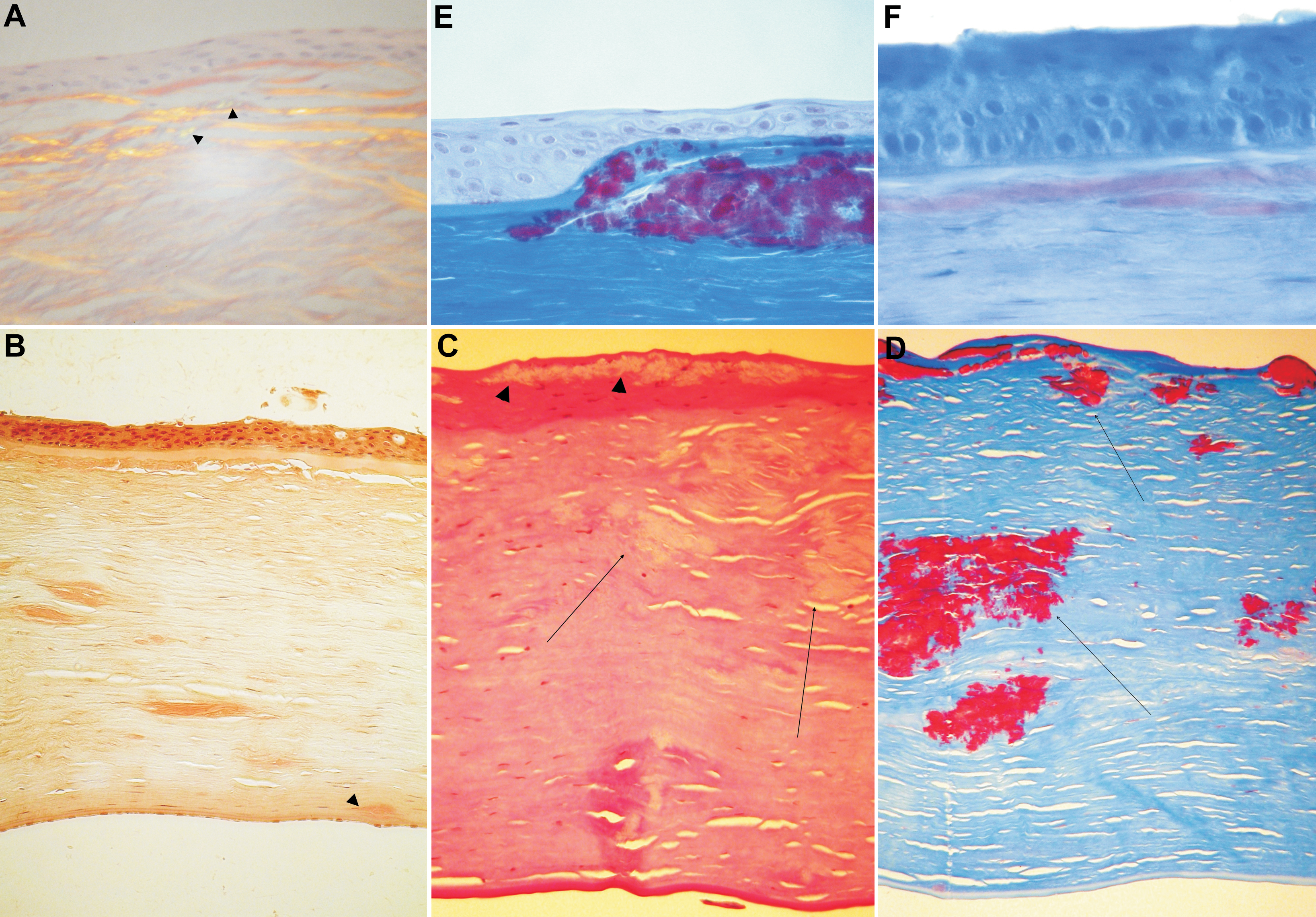
 Figure 3
of Nowińska, Mol Vis 2011; 17:2333-2342.
Figure 3
of Nowińska, Mol Vis 2011; 17:2333-2342.