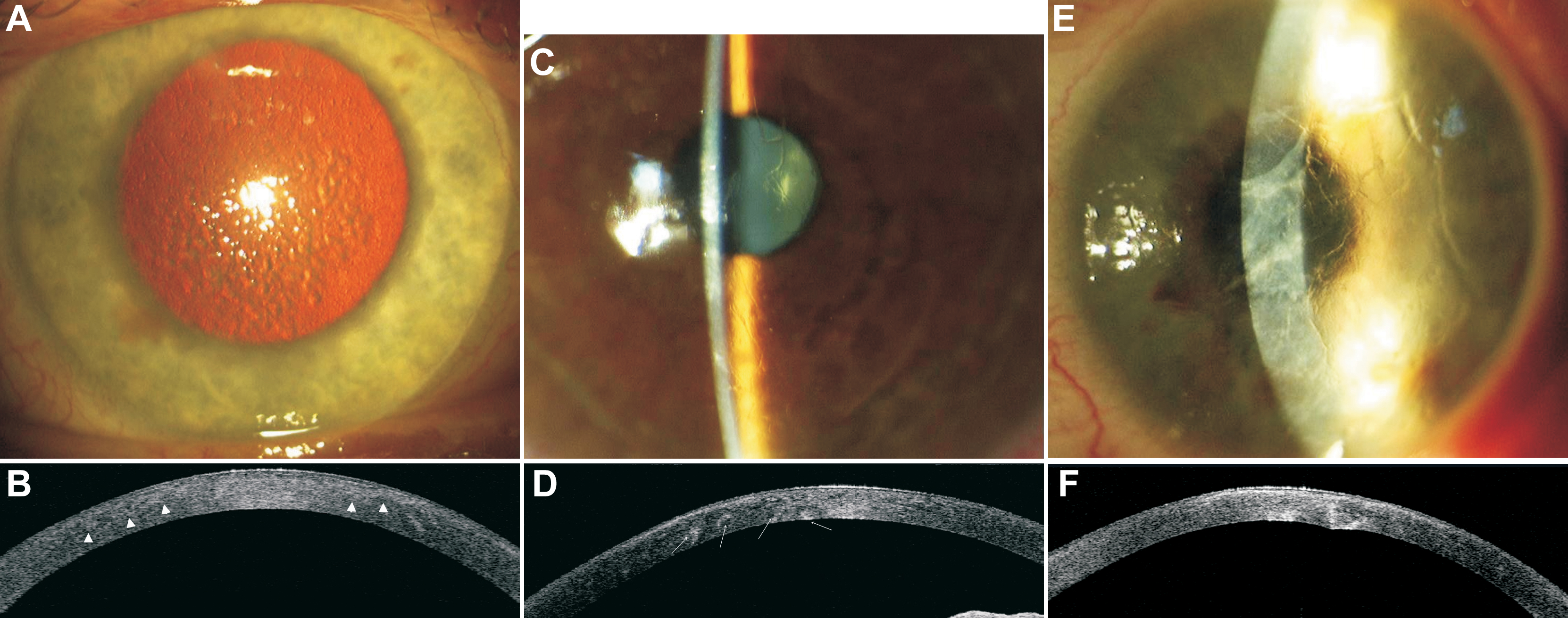
Figure 2. Representative images of
slit-lamp photographs and 1310 nm time-domain optical coherence
tomography scans of patients with lattice corneal dystrophy type
I (family F1); lattice corneal dystrophy variants (families F2
and F7). There is a noticeable phenotypic heterogeneity between
corneal morphology of lattice corneal dystrophy variants caring
the same H626R mutation.
A: Male patient (F1; 37 years).
Slit-lamp retroillumination photograph showing diffuse multiple
lattice lines. LCDI/R124C mutation.
B: Male patient (F1;
37 years). High-resolution corneal scan – 1310 nm time. domain
OCT. There is a diffuse border between the anterior part of
increased reflectivity and normal corneal stroma (arrowheads).
The areas of increased stromal reflectivity correspond with
corneal opacities. LCDI/R124C mutation.
C: Female
patient (F2; 45 years). Slit-lamp photograph. Delicate, fragile,
rare lattice lines located centrally. LCD variant/ H626
mutation.
D: Female patient (F2; 45 years).
High-resolution corneal scan – 1310 nm time. domain OCT.
Opacities with increased reflectivity visible through the whole
depth of the cornea. Some of the opacities are located in the
posterior corneal part (arrows). LCD variant/H626 mutation.
E:
Female patient (F7; 48 years). Slit-lamp photograph. Thick,
distinct lines accompanied by stromal haze extended from limbus
to limbus. LCD variant/H626 mutation. Note the distinct
heterogeneity compared to
Figure 2C.
F:
Female patient (F7; 48 years). High-resolution corneal scan –
1310 nm time. domain OCT. Opacities with increased reflectivity
located mainly in the posterior corneal part causing distortion
of the posterior corneal surface. LCD variant/H626 mutation.
 Figure 2
of Nowińska, Mol Vis 2011; 17:2333-2342.
Figure 2
of Nowińska, Mol Vis 2011; 17:2333-2342.