Figure 5. Electron micrograph of tree shrew and human cornea fixed in 2.5% glutaraldehhyde containing cuprolinic blue and embedded in
spurr resin. A: Electron micrograph of proteoglycans of tree shrew stroma. B: Digital image obtained after processing image shown in A, showing variable area distribution of PGs. C: Electron micrograph of proteoglycans of human stroma. D: Digital image obtained after processing image shown in C, showing variable area distribution of PGs. PG=Proteoglycan; Proteoglycans color coding: Red=50–350 nm2, Green=350–650 nm2, Blue=650–950 nm2, Yellow=950–1,250 nm2, Aqua=1,250–1,550 nm2, Pink=1,550–1,850 nm2, and Brown=1,850–2,150 nm2.
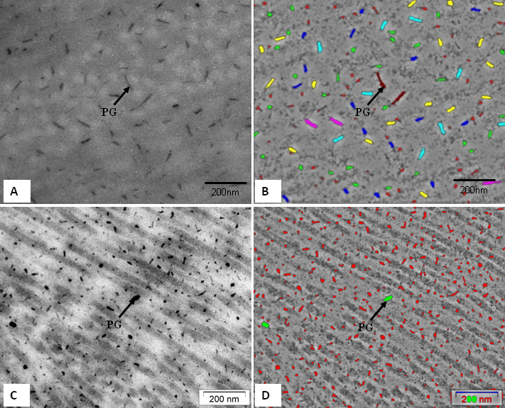
 Figure 5 of
Almubrad, Mol Vis 2011; 17:2283-2291.
Figure 5 of
Almubrad, Mol Vis 2011; 17:2283-2291.