Figure 3. Electron micrograph of tree shrew cornea fixed in 2.5% glutaraldehyde containing cuprolinic blue and embedded in spurr resin.
A: In the middle stroma, parallel running lamellae containing a keratocyte. B and C: Lamella containing orderly, packed collagen fibrils and proteoglycans. D: In cross-section, collagen fibrils exhibiting tiny particles, some of which are of high electron density. E: Pre-Descemet's stroma containing very fine fibrils and large PGs around the collagen fibrils. Fibrillar structures are present
throughout the Descemet's membrane. F: Part of the posterior cornea, showing a banded Descemet's membrane and an endothelium containing a nucleus; also a prominent
endoplasmic reticulum. CF=Collagen fibrils, DM=Descemet's membrane, EN=endothelium, F=Fine fibril, G=tiny particles, KR=Keratocytes,
L=Lamella, PG=Proteoglycan, and S=Stroma.
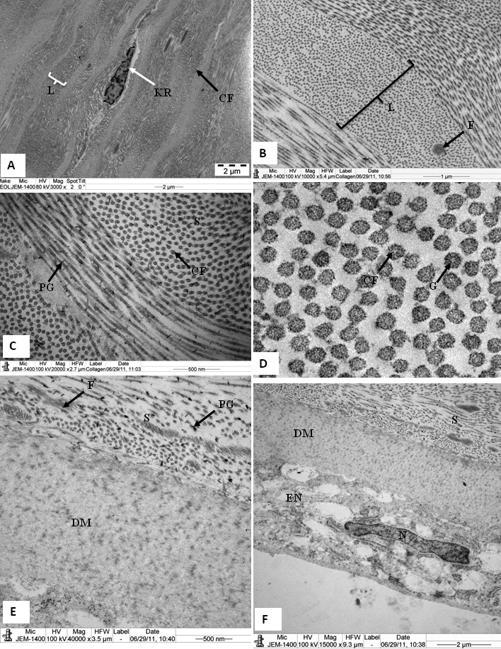
 Figure 3 of
Almubrad, Mol Vis 2011; 17:2283-2291.
Figure 3 of
Almubrad, Mol Vis 2011; 17:2283-2291.