Figure 2. Electron micrograph of tree shrew and human cornea fixed in 2.5% glutaraldehyde containing cuprolinic blue and embedded in
spurr resin. A: Basal epithelial cells are columnar and contain large nuclei. B: Prominent cytoplasmic filaments in basal epithelial cells. C: Basal epithelial cells attached by hemidesmosomes to a basement membrane followed by a Bowman’s layer consisting of dense,
randomly arranged collagen fibrils. D: Non linear, random distribution of collagen fibrils (curved arrowhead) present in the anterior stroma just below the Bowman's
layer. Some of the collagen run across the longitudinally-running collagen fibrils (arrowhead). E: Lamellae are interlacing (arrowhead) in the anterior stroma of the tree shrew. F: Lamellae are interlacing (arrowhead) in the anterior stroma of the normal human cornea. B=Bowman’s layer, BM=Basement membrane,
CF=Collagen fibrils, E=Epithelium, H=Hemidesmosomes, and MF=Cytoplasmic filaments.
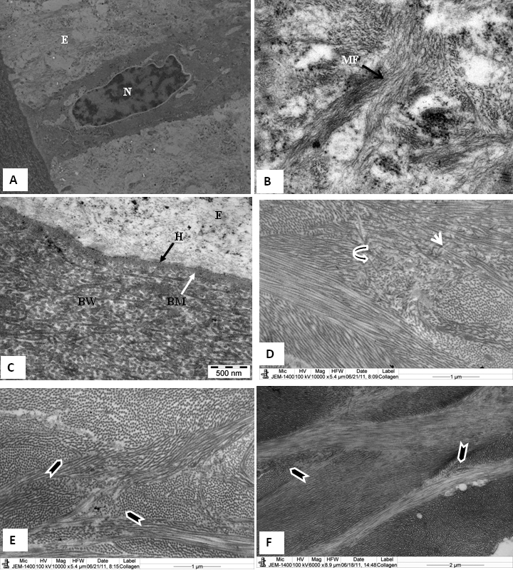
 Figure 2 of
Almubrad, Mol Vis 2011; 17:2283-2291.
Figure 2 of
Almubrad, Mol Vis 2011; 17:2283-2291.