Figure 1. Light micrograph of tree shrew cornea. A: Tree shrew cornea consisting of epithelium, Bowman’s layer, stroma, Descemet’s membrane, and endothelium. Note the structure
of the cornea is very similar to the human cornea and shows 5 layers. B: Part of the tree shrew cornea showing squamous cells, wing cells, basal epithelial cells and Bowman’s layer. C: Part of the corneal stroma containing keratocytes. D: Part of the cornea showing Descemet’s membrane and endothelium. B=Bowman’s layer, E=Epithelium, DM=Descemet’s membrane,
KR=Keratocyte, EN=endothelium, and S=Stroma.
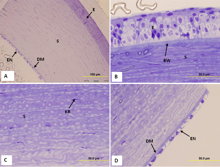
 Figure 1 of
Almubrad, Mol Vis 2011; 17:2283-2291.
Figure 1 of
Almubrad, Mol Vis 2011; 17:2283-2291.