Figure 6. Fundus signs of proband. Color photographs and posterior segment optical coherence tomography of proband 2. The color photographs
show significant subretinal fluid in both eyes under the central vitelliform lesions, confirmed on optical coherence tomography.
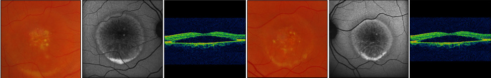
 Figure 6 of
Low, Mol Vis 2011; 17:2272-2282.
Figure 6 of
Low, Mol Vis 2011; 17:2272-2282.