Figure 3. Anterior segment optical
coherence tomography findings for family 1. A-D:
Right eyes of individuals II:1 to II:4 respectively. E-H:
Left eyes of individuals II:1 to II:4 respectively. The
unaffected sister (II:2) shown in row 2 had closed angles on
anterior segment (AS)-OCT. Slit openings were observed for
individuals II:1 and II:3 on AS-OCT but their angles were closed
on gonioscopy. The proband shown in row 4 had the shallowest
anterior chamber depths. H: This shows a thickened
cornea from aphakia and corneal decompensation from previous
surgery in the proband.
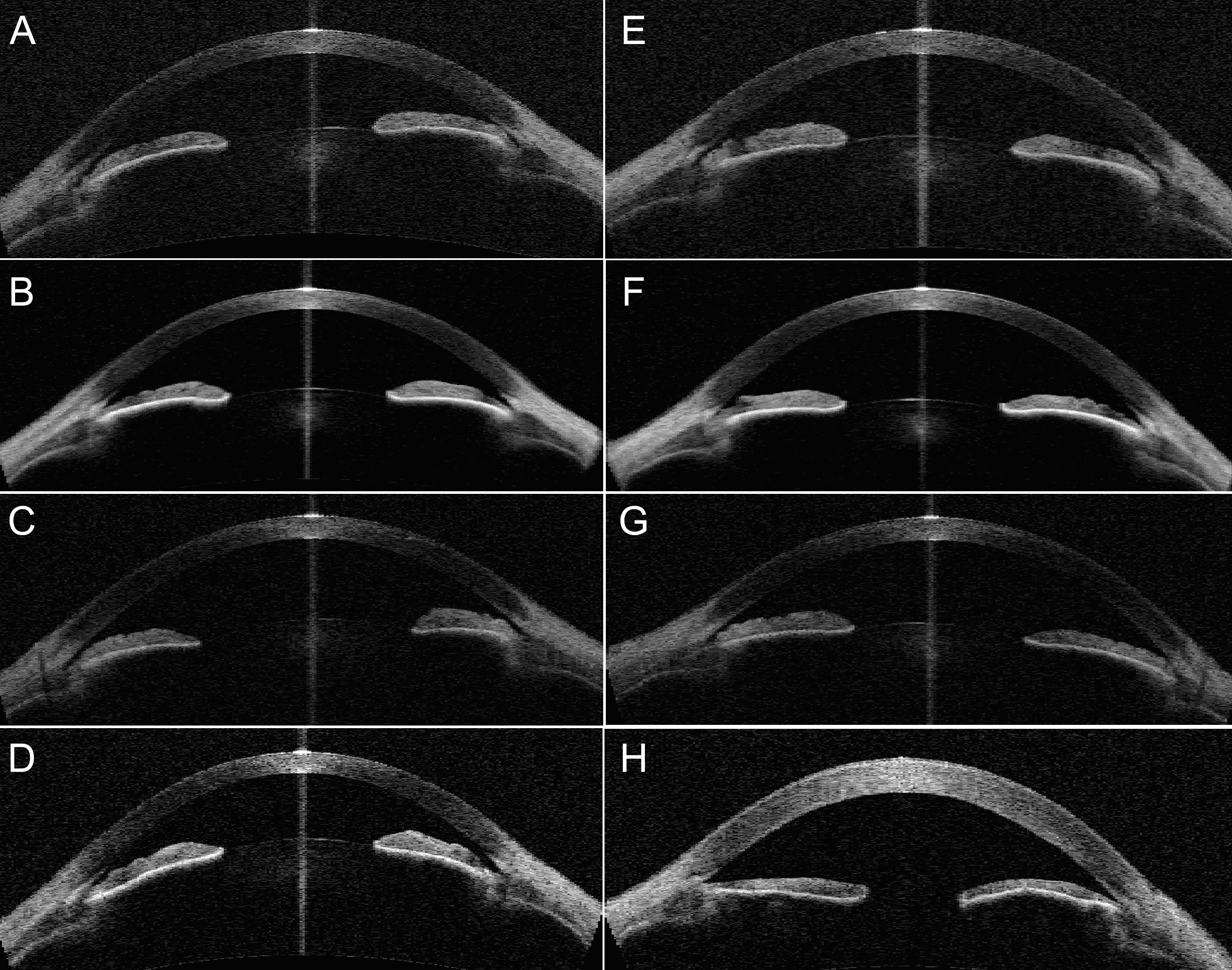
 Figure 3
of Low, Mol Vis 2011; 17:2272-2282.
Figure 3
of Low, Mol Vis 2011; 17:2272-2282.