Figure 2. Fundus signs of affected
siblings in family 1. Fundus photographs, autofluorescence, and
posterior segment optical coherence tomography (OCT) images of
family 1. A: Individual II:1 (affected sister) had a
completely normal left posterior segment examination but
significant retinal pigment epithelium (RPE) layer thickening on
the right. B: Individual II:3 (affected brother) had
disruption of the inner segment: outer segment interface. C:
Individual II:4 (proband) had exudative vitelliform lesions,
demonstrated in the right eye.
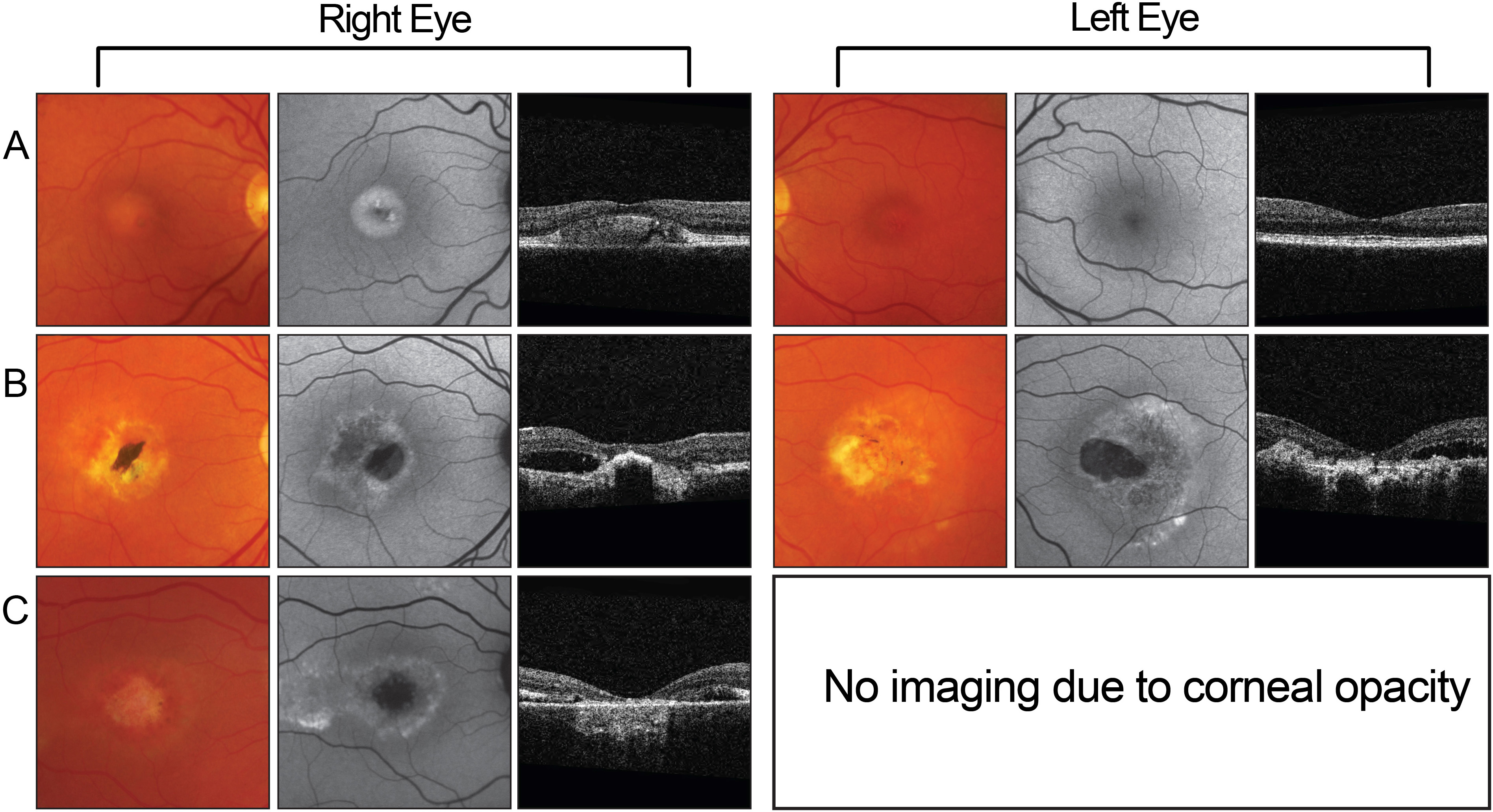
 Figure 2
of Low, Mol Vis 2011; 17:2272-2282.
Figure 2
of Low, Mol Vis 2011; 17:2272-2282.