Figure 3. Immunofluorescent staining
of S100A2, A6, A8, A10, and A11 proteins (in green) in normal
human corneal, limbal and SCC epithelia. The nucleus was
counter-stained blue with DAPI. Except for the pictures on
S100A10 staining, which were taken at 400×, all others were
taken at 200× magnification originally.
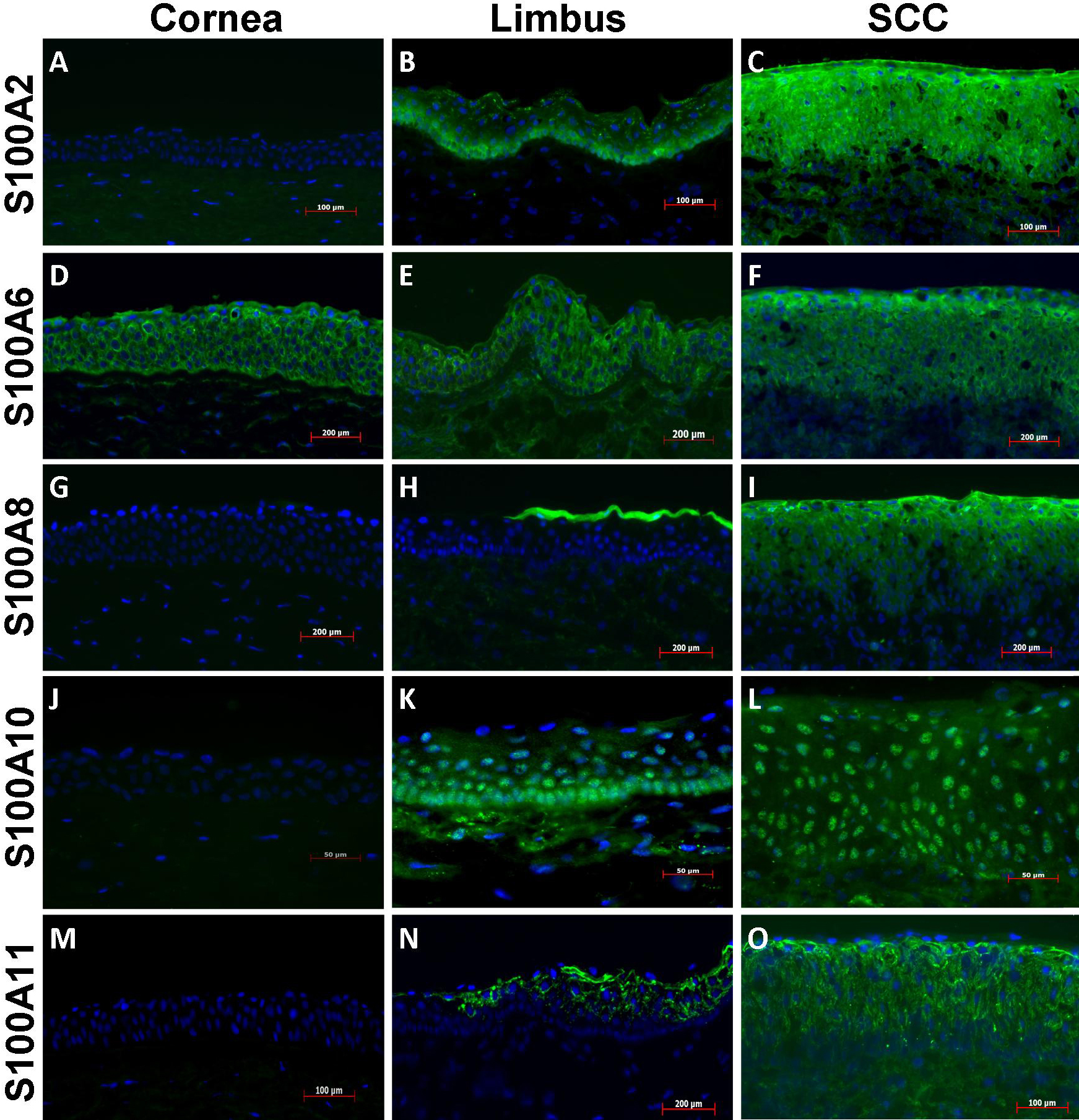
 Figure 3
of Li, Mol Vis 2011; 17:2263-2271.
Figure 3
of Li, Mol Vis 2011; 17:2263-2271.