Figure 2. Morphological features and
cytokeratin protein expression in SCC tissue. A and B:
H&E staining of SCC tissue. A: Notice the thickening
of the epithelium as demarcated by the white dotted line, the
breach of the basement membrane and massive leukocytes
infiltration (white arrows). B: The epithelial cell
invasion of the stroma (marked by the dotted white lines). The
keratin pearls formed by necrotic epithelial cells (white
arrow). The invading cells showed typical metaplastic appearance
with enlarged nucleus and cell volume. The original pictures
were taken at 100× magnification. C and D:
Immunofluorescent staining of cytokeratin proteins (green) in
SCC tissue. C: CK14 antibody stained the full thickness
of the epithelium. The signal was more intense along the basal
cell layer. D: CK4 staining of the SCC epithelium except
for the basal layer. E: Weak CK3/12 staining of the SCC
epithelium. The nucleus was counter-stained blue with DAPI. The
original pictures were taken at 200× magnification.
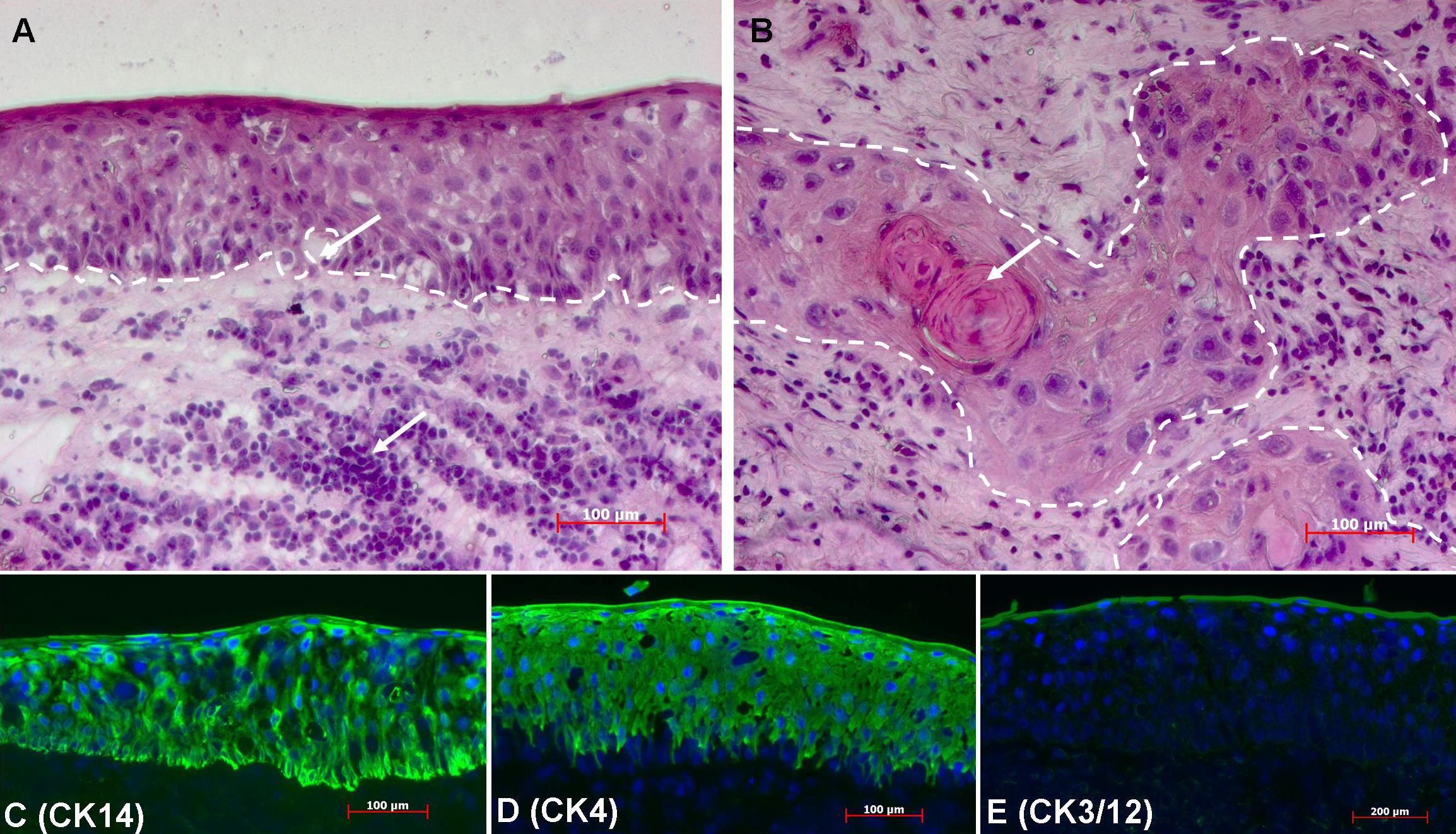
 Figure 2
of Li, Mol Vis 2011; 17:2263-2271.
Figure 2
of Li, Mol Vis 2011; 17:2263-2271.