Figure 1. A: Fundus photography of patient 1 (P1) demonstrating bilateral cystoid-like macular lesions. There are no pigmentary changes.
B: Optical coherence tomography (OCT) scans through the macular region of P1 (upper part) and P2 (lower part). There is loss
of foveal contour (asterisks) and thinning of the hyporeflective layer representing the photoreceptors in the macular region
(white arrows). P1 and P2 scans are off-center, but foveas can be seen. C: Reference areas within an OCT through the macular region of a normal patient. GCL, Ganglion cell layer; INL, inner nuclear
layer; IPL, inner plexiform layer; NFL, nerve fiber layer; OPL, outer plexiform layer; RPE, retinal pigment epithelium.
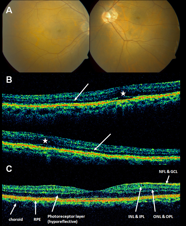
 Figure 1 of
Kinori, Mol Vis 2011; 17:2241-2247.
Figure 1 of
Kinori, Mol Vis 2011; 17:2241-2247.