Figure 4. The cellular localization of PKR associated protein X (RAX) in the retinas of streptozotocin-induced diabetic rats by immunofluorescence.
The retinas of diabetic rats show a strong cytoplasmic expression of RAX at 6 days (B) after STZ injection when compared to normal rats (A). The RAX expression in the retinas of diabetic animals at 28 days (C) and 35 days (D) after STZ injection was strongly reduced to the level of normal rats (A). The nuclei of cells (E, F, G, and H) were stained with the 4',6-diamidino-2-phenylindole (DAPI, blue) and the negative controls sections (I, J, K, and L) were incubated without the anti-RAX antibody. The negative control sections (incubated without the anti-RAX antibody) of
the normal (I) and diabetic rat retinas at 6, 28, and 35 days are J, K, and L, respectively. Arrows indicate that RAX protein was expressed in the ganglion cell layer and the inner nuclear layer of the
retina. The original magnification was 400×. The scale bar represents 50 μm. Abbreviations are as follows: GCL represents
ganglion cell layer; INL represents inner nuclear layer; ONL represents outer nuclear layer; SRS represents subretinal space.
The results shown in this figure are representative of three replicates.
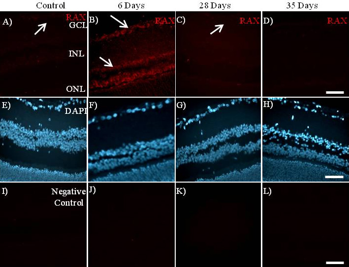
 Figure 4 of
Silva, Mol Vis 2011; 17:2228-2240.
Figure 4 of
Silva, Mol Vis 2011; 17:2228-2240.