Figure 1. The cellular localization of microRNA-29b in the retina of streptozotocin (STZ)-induced diabetic rats by in situ hybridization.
The miR-29b signal was weak in the retina of diabetic rats at 6 days (B), but strong at 28 days (C) and 35 days (D) after STZ injection when compared to normal rats (A). The negative controls (E, F, G, and H) were obtained with sections incubated in the absence of anti-digoxigenin (DIG)-labeled probe. The positive control sections
(U6 RNA) of the normal and diabetic rat retinas at 6, 28, and 35 days are I, J, K, and L, respectively. Arrows indicate that miR-29b was expressed in the ganglion cell layer and the inner nuclear layer of the retinas. Original magnification was 400×. The
scale bar represents 50 μm. Abbreviations are as follows: GCL represents ganglion cell layer; INL represents inner nuclear
layer; ONL represents outer nuclear layer; OS represents photoreceptor outer segments. The results shown in this figure are
representative of three replicates.
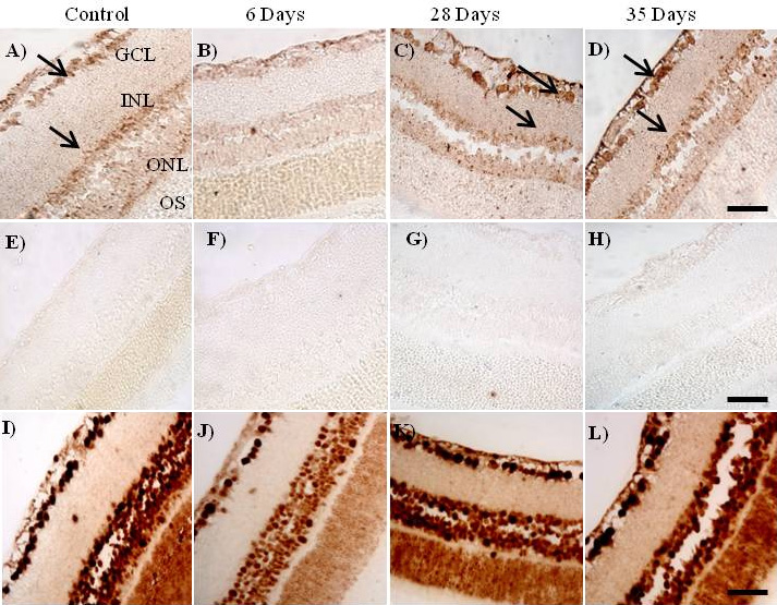
 Figure 1 of
Silva, Mol Vis 2011; 17:2228-2240.
Figure 1 of
Silva, Mol Vis 2011; 17:2228-2240.