Figure 7. CD45, actin, and nuclei
staining. CD45 (cyan), Actin (green) and Nuclei (red) staining
of corneas from 7 (A) and 10 month old (B) Ctns−/−
mice. Panel A shows few CD45 positive stained cells in
the limbus of 7 month old mice while panel B shows
abundant CD45 stained cells in the limbus and central cornea of
10 month old Ctns−/− mice.
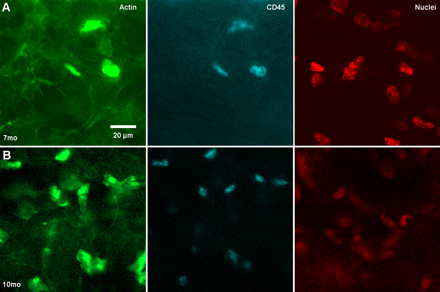
 Figure 7
of Simpson, Mol Vis 2011; 17:2212-2220.
Figure 7
of Simpson, Mol Vis 2011; 17:2212-2220.