Figure 5. Corneal staining with actin and nuclei staining. A: Actin (Green) and nuclei (red) staining of cornea in normal C57Bl/6 mouse using confocal microscopy. We observed normal
keratocytes cell bodies with dendritic morphology that stained in the periphery for actin. B: Actin and nuclei staining of corneas from Ctns−/− mice at 5 months of age showing few inflammatory cells (arrow). Inflammatory cell infiltration appeared to increase in the
10th (C, arrows) and 12th (D, arrows) month of age with keratocyte dropout (asterisk).
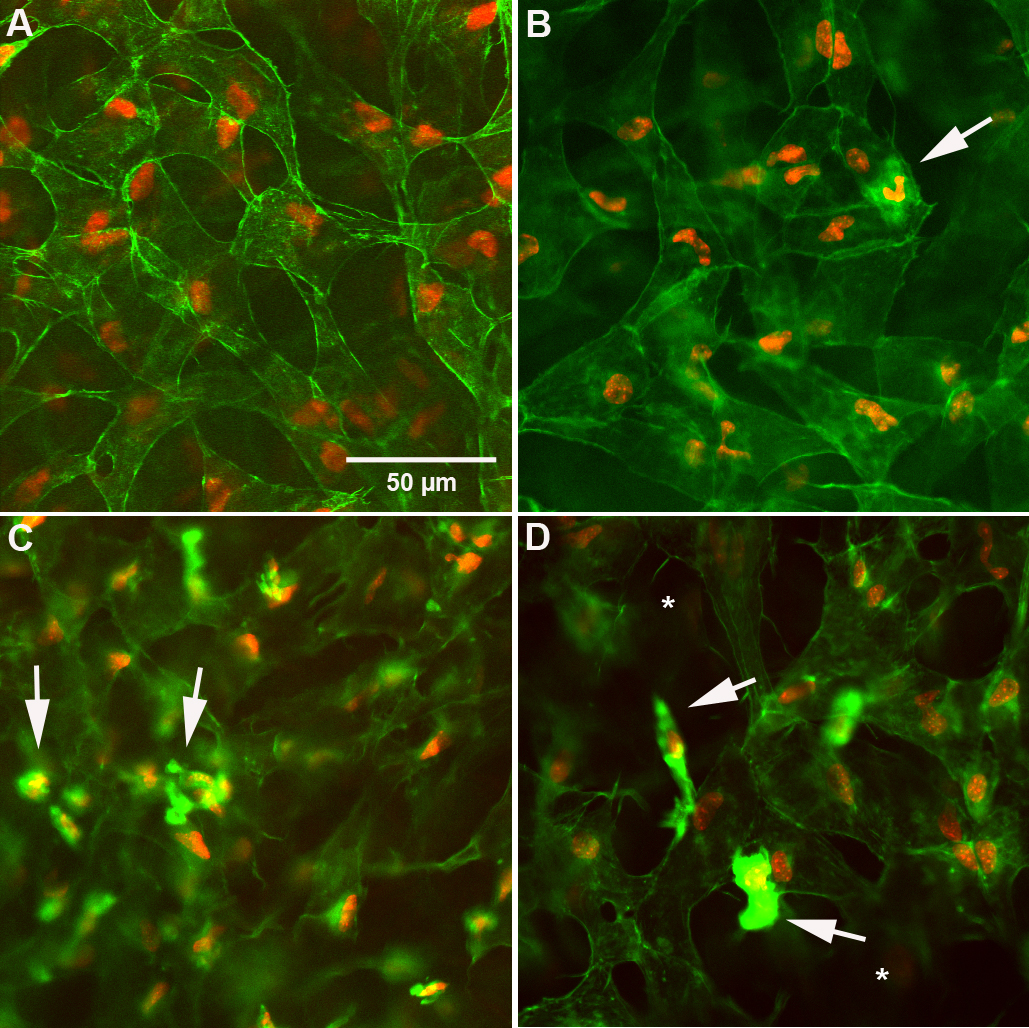
 Figure 5 of
Simpson, Mol Vis 2011; 17:2212-2220.
Figure 5 of
Simpson, Mol Vis 2011; 17:2212-2220.