Figure 3 of
Simpson, Mol Vis 2011; 17:2212-2220.
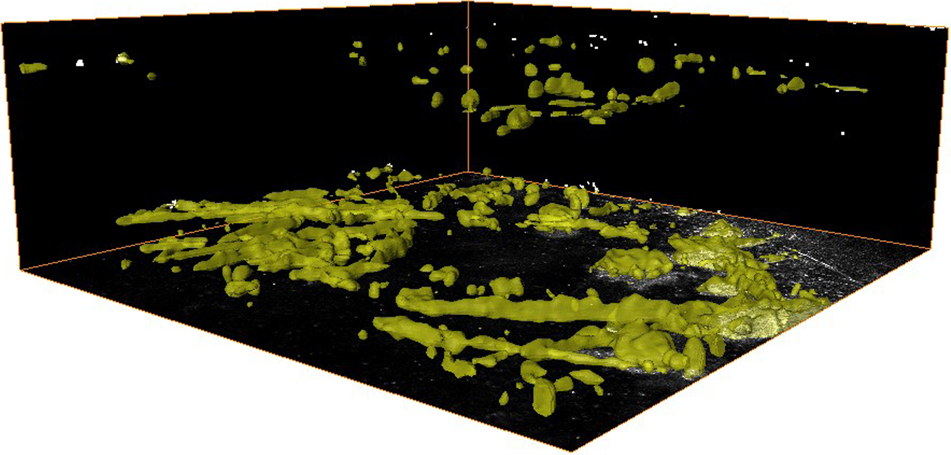
Figure 3.
3-D distribution of crystals within the cornea of a 7month old
Ctns
−/−
mouse. Crystal volume was segmented from the 3-D in vivo CM scan by thresholding the images to contain just the crystal volume.
 Figure 3 of
Simpson, Mol Vis 2011; 17:2212-2220.
Figure 3 of
Simpson, Mol Vis 2011; 17:2212-2220.