Figure 2. Confocal imaging time
course. Confocal images of Ctns−/− corneas at
3 months (A), 7 months (B), 10 months (C),
and 12 months of age (D). Each panel shows a xy (upper)
and xz (lower) slice through a 3-D stack. Cystine crystals were
identified as small, 20 µm long, needle-like crystals in the
peripheral and central cornea. Cystine crystals were first
detected in 3 month old mice (A, arrow) and gradually
increased in density with age up to 7 months (B). Older Ctns−/−
mice (10 and 12 month old) developed aggregates of brightly
reflecting material, presumably cystine, within the central
cornea (arrowheads; C and D, respectively).
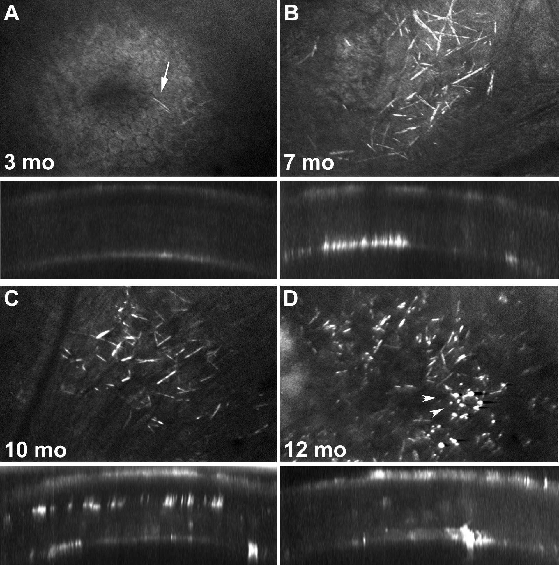
 Figure 2
of Simpson, Mol Vis 2011; 17:2212-2220.
Figure 2
of Simpson, Mol Vis 2011; 17:2212-2220.