Figure 5. En face confocal immunofluorescence microscopy of Schlemm’s canal inner wall. Shown is labeling of the inner wall of SC (viewed
from luminal side) for integrin α6 (A, green), laminin α5 (B, red), CD31 (C, blue). Panel D is a merged image of all three proteins plus nuclei (brown). Shown is representative image from a human donor eye of 6 total
eyes that were examined.
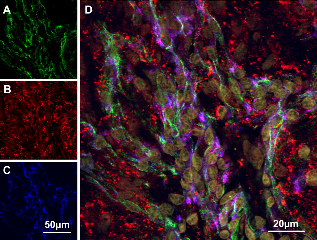
 Figure 5 of
VanderWyst, Mol Vis 2011; 17:199-209.
Figure 5 of
VanderWyst, Mol Vis 2011; 17:199-209.