Figure 4. Integrin α6 subunit expression by Schlemm’s canal cells. A: Confocal immunofluorescence microscopy of outflow tissues including Schlemm’s canal (SC) from human donor eyes showing α6
integrin (INTG) levels. Shown is representative image from eye of individual human donor of 3 donor eyes that were examined.
For comparison, a scleral vessel is indicated (*). B: western blot analysis of different human SC and trabecular meshwork (TM) cell strains checking for α6 integrin levels. Cell
lysates were first subjected to immunoprecipition using integrin α6-specific IgGs, followed by SDS–PAGE and western blotting
using IgGs that recognize both α3 and α6 INTG.
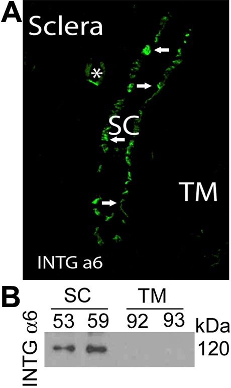
 Figure 4 of
VanderWyst, Mol Vis 2011; 17:199-209.
Figure 4 of
VanderWyst, Mol Vis 2011; 17:199-209.