Figure 3. Vascular endothelial integrins in human conventional outflow pathway and cultured cells. A: Confocal immunofluorescence microscopy of outflow tissues from human, post-mortem eyes examining integrin (INTG) subunit
levels. Shown are results obtained from one eye from one individual donor of three total that were examined. Background fluorescence
in the absence of primary antibodies is shown (2°). B: western blot analysis of integrin levels by different human Schlemm’s canal (SC) and trabecular meshwork (TM) cell strains
in culture.
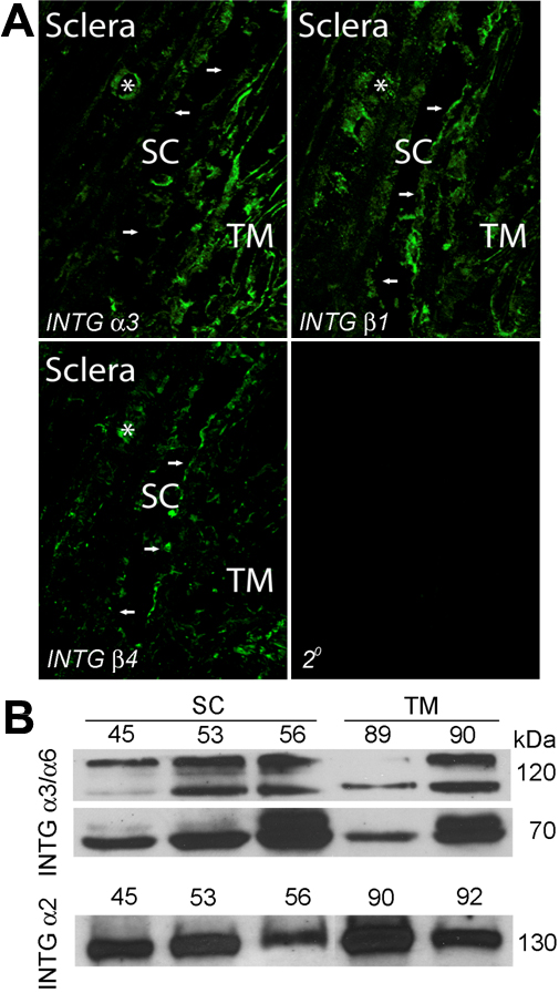
 Figure 3 of
VanderWyst, Mol Vis 2011; 17:199-209.
Figure 3 of
VanderWyst, Mol Vis 2011; 17:199-209.