Figure 2. Laminins-332 and −511 in human conventional outflow pathway and cultured cells. A: Confocal immunofluorescence microscopy of endothelial laminins (LM) in radial sections through human conventional outflow
tissues. Shown are representative images taken from one human donor eye of three that were examined. B: western blot analysis of endothelial laminin levels by different strains of cultured SC and TM cell monolayers isolated
from different individual eye donors. Samples for laminin α5 blots were obtained after cell lysates were first immunoprecipitated
with anti-laminin γ1 IgGs.
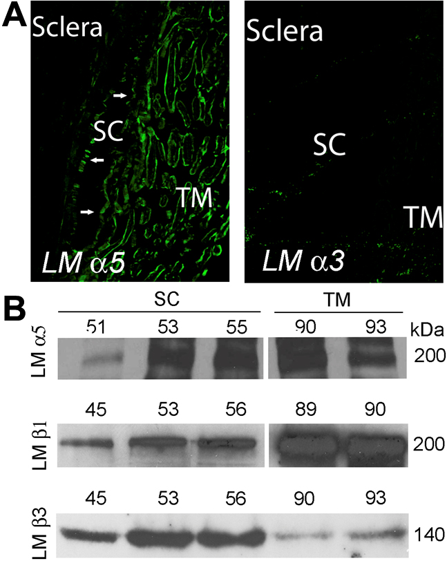
 Figure 2 of
VanderWyst, Mol Vis 2011; 17:199-209.
Figure 2 of
VanderWyst, Mol Vis 2011; 17:199-209.