Figure 1. Collagens in human conventional outflow pathway and cultured cells. A: Confocal immunofluorescence microscopy of outflow tissues from cadaveric human eyes examining the expression levels of collagen
(CN) I, III, and IV. Vascular endothelial cadherin (VE-cad) labeling was used as positive control for integrity of tissue
antigens and localization of Schlemm’s canal (SC). Shown are representative images taken from section of one donor eye of
3 examined. B: Western blot analysis of collagen levels in different strains of cultured human trabecular meshwork (TM) and SC cell monolayers
isolated from different individual eye donors. Anti-collagen I IgGs recognized multiple bands between 140 and 40 kDa, corresponding
to post-translational modifications.
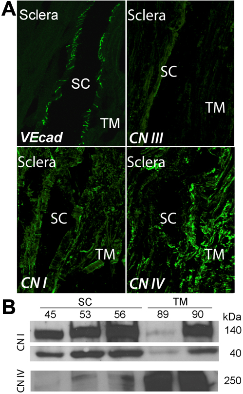
 Figure 1 of
VanderWyst, Mol Vis 2011; 17:199-209.
Figure 1 of
VanderWyst, Mol Vis 2011; 17:199-209.