Figure 2. Sequence analysis of CRYGD. A: Sequence of a member without the polymorphism. B: Heterozygous polymorphism detected in exon 3 of CRYGD (individuals II-15, III-22, and IV-7). C: Multiple sequence alignment of the CRYGD protein (codons 120–145) in different species, demonstrating that residue 134 is
highly conserved. D: Slit-lamp photograph, showing pulvurulent cataract in individual III-22 (left) and lamellar cataract in individual IV-7
(right).
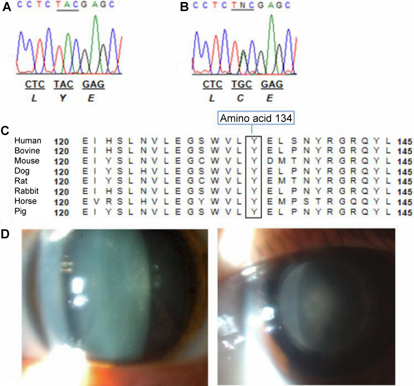
 Figure 2 of
de Figueirêdo, Mol Vis 2011; 17:2207-2211.
Figure 2 of
de Figueirêdo, Mol Vis 2011; 17:2207-2211.