Figure 2. Putative LESC marker expression patterns in normal and diabetic ex vivo limbus. Note a dramatic decrease in staining intensity
and the number of positive basal epithelial cells for ABCG2 and ΔNp63α in the diabetic limbus. ΔNp63α was revealed with two
different antibodies (Santa Cruz, SC) and Pellegrini (P) with the same result. e, epithelium, s, stroma. Bar=30 μm.
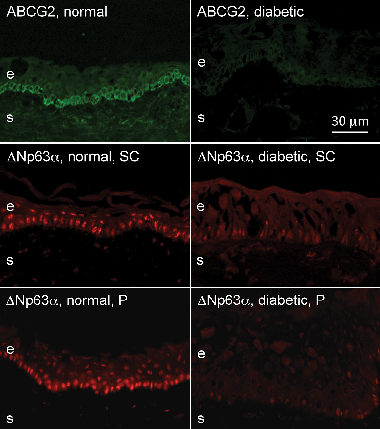
 Figure 2 of
Saghizadeh, Mol Vis 2011; 17:2177-2190.
Figure 2 of
Saghizadeh, Mol Vis 2011; 17:2177-2190.