Figure 4. The histopathological changes and decrease in Bax expression in p50-deficient (O) mice with chronic administration of tacrolimus:
A: Light micrographs of cross sections derived from the un-treated wild type (WT), and p50-deficient (KO) mice at 10 month
of ages, and the age-matched KO mice treated by chronic administration of tacrolimus, lomerizine or memantine. Although the
histopathological results show obvious excavation of the optic nerve head and RGC loss in the un-treated KO mice, no cupping
is observed in the KO mice treated by chronic administration of tacrolimus, lomerizine or memantine. Serious losses in surviving
RGCs are not detected in the KO mice treated with tacrolimus. Original magnification; (upper panels, low magnification 5×;
lower panels, high magnification 20×). B: Azan staining; although the increase in collagenous fibers (blue color staining) caused by the axonal loss was clearly detected
in optic nerve sections of KO mice, the chronic administration of tacrolimus prevented the increase (upper panel: original
magnification; 40×). Electron microscopic examinations were performed for detection of morphological abnormalities of cross-sectioned
optic nerve axons (lower panel). The KO mice exhibit an expanding area of each axon as well as decreasing number of axons
in comparison with age-matched WT mice (lower panel). The area of connective tissue was significantly increased in KO mice
(lower panel). However, these changes were not observed in the KO mice with the chronic administration of tacrolimus (lower
panel). Scale bar=10 μm. C: The numbers of axons were counted from 10 fields of identical size (18 μm×18 μm). The Bar graph shows a significant decrease
at 10 months of age in KO mice compared to parental mice (WT) and the KO mice treated with tacrolimus (Tac.). Data are the
mean±SEM (each 5). Data were analyzed by the Mann–Whitney U-test (**p<0.0001, *p<0.001). D: Immunohistochemical staining with a-Bax antibody (clone 6A7) or normal mouse serum plus tissue sections from 9-month-old
wild type mice (WT) and p50-deficient (KO) mice with and without chronic administration of tacrolimus. Bax expression was
observed in the GCL of KO mice, but not age-matched KO mice treated with tacrolimus (Tac.) or WT mice. GCL=ganglion cell layer,
IPL=inner plexiform layer. Green; Bax, Red; PI. Original magnification; 100×.
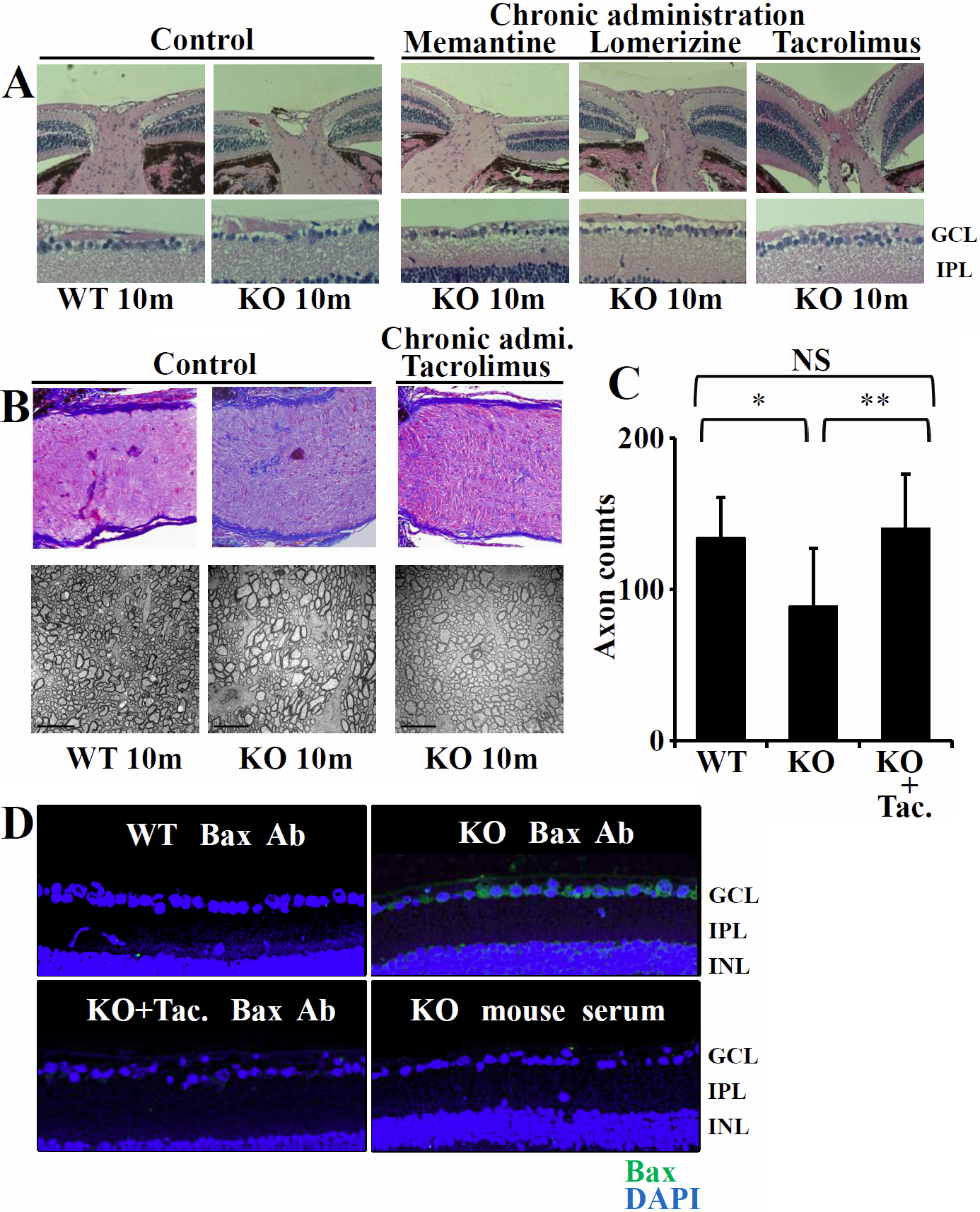
 Figure 4 of
Nakamura-Yanagidaira, Mol Vis 2011; 17:2157-2170.
Figure 4 of
Nakamura-Yanagidaira, Mol Vis 2011; 17:2157-2170.