Figure 5. SDF-1α-induced VEGF production by peritoneal macrophages. A: CXCR4 expression on F4/80-positive murine peritoneal macrophages was determined by a flow cytometric analysis. Purified
mononuclear cells were stained with rat anti-mouse F4/80 mAb and rabbit anti-CXCR4 Abs (filled histogram) or rat anti-mouse
F4/80 mAb and non-immunized rabbit IgG (open heavy-lined histogram) as a control followed by staining with FITC-conjugated
goat anti-rabbit IgG and PE-conjugated swine anti-rat IgG. A representative result from three independent experiments is shown.
B: Peritoneal macrophages from WT mice were incubated with the indicated concentrations of SDF-1α for 12 h. Quantitative RT–PCR
was performed on total RNAs extracted from the macrophages as described in Methods. Representative results from 3 independent
experiments are shown here. C: VEGF mRNA levels were determined and normalized to Actb mRNA levels. Each value represents the mean and SEM (n=3). #, p<0.01 compared with untreated. D: VEGF protein expression was detected by immunocytochemical analysis using anti-VEGF Abs as described in Methods. Representative
results from three independent experiments are shown. Original magnification, 400×. Scale bar, 50 μm.
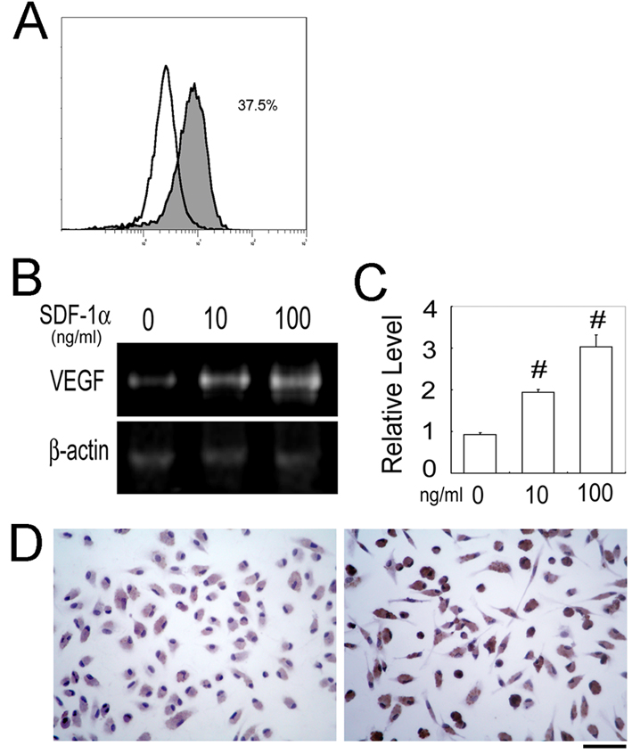
 Figure 5 of
Liu, Mol Vis 2011; 17:2129-2138.
Figure 5 of
Liu, Mol Vis 2011; 17:2129-2138.