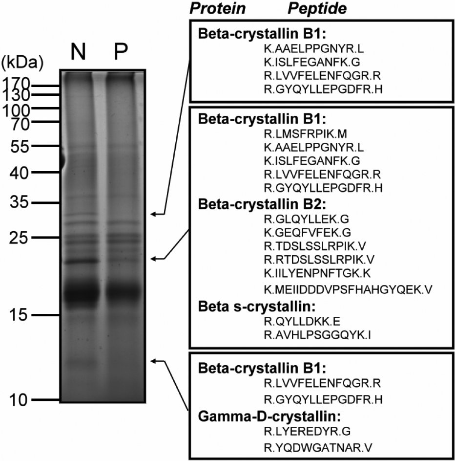
Figure 1. Comparative analysis of normal and cataractous human lens proteins by SDS–PAGE followed by LC-nanoESI-MS/MS. As shown in the
left panel, a total of 10 μg lens proteins derived from normal (N) and cataractous (P) eye lenses were resolved with 12.5%
SDS–PAGE and stained with Coomassie brilliant blue R-250. In the right panel, protein and peptide bands with different expression
levels identified by LC-nanoESI-MS/MS were indicated by arrows. In comparison with normal human lens proteome, four crystallin
proteins, β-crystallin B1, β-crystallin B2, βs-crystallin, and γD-crystallin in the cataract lens were found to significantly
decrease in expression levels as compared to normal lens.

 Figure 1 of
Huang, Mol Vis 2011; 17:186-198.
  Figure 1 of
Huang, Mol Vis 2011; 17:186-198.