Figure 2. Ultrasound and MRI findings in III:1 and II:1. The father’s (II:1) Doppler ultrasonographic examination. This demonstrates
A: a normal right eye with the optic nerve and arteria centralis retinae; B: a left microphthalmic heterogeneous eye without the optic nerve visible. C, D: The son’s (III:1) Doppler ultrasonographic examination demonstrating the absence of both optic nerves and corresponding
vascularization, but the presence of posterior ciliary vessels (C and D for right and left eye, respectively). The vessels are represented in color. The Doppler examination is represented in the
boxes. E: II:1’s axial T2-weighted image in MRI demonstrated a normal right eye and lens and a left microphthalmic eye with thick
sclera. F: III:1’s axial T2-weighted image in MRI showed an almost normal morphology of both eyes but the absence of optic nerves (with
some remnants of dural sheath), chiasms, and tracts. G-H: Coronal T1-weighted MRI images in the midorbital (G) and intracranial (H) planes. This shows the complete lack of both orbital nerves in the son, III:1.
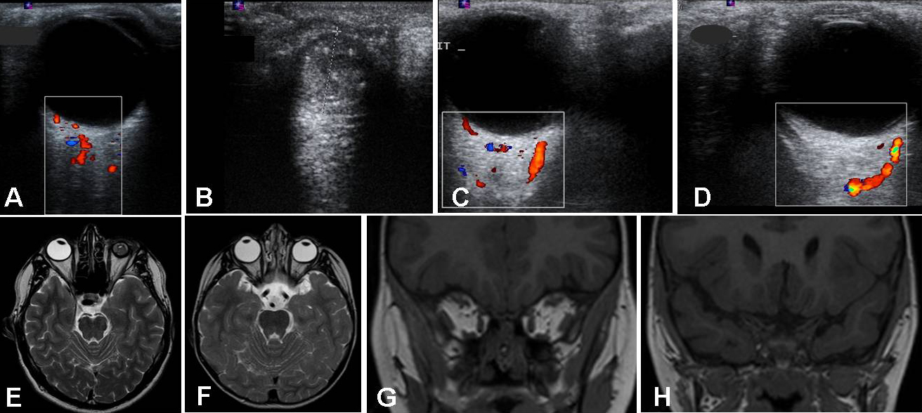
 Figure 2 of
Meire, Mol Vis 2011; 17:2072-2079.
Figure 2 of
Meire, Mol Vis 2011; 17:2072-2079.