Figure 1. Clinical pictures of III:2, III:1 and II:1. A: A picture of III:2 with mild bilateral microphthalmia with 10.5 mm corneal diameters and atypical coloboma of the iris in
the right eye. B: A picture of twin brother III:1 with bilateral microphthalmia with 9 mm corneal diameters and scleralization of the inferior
cornea. C-D: Fundus picture of III:1 showing the absence of the optic nerve, dysplastic retinae, and a few retinal vessels. E: A picture of the father’s (II:1) left eye, showing unilateral left microphthalmos (corneal diameter of 7 mm) with a vascularized
cornea, impairing the view to the anterior segment and to the fundus.
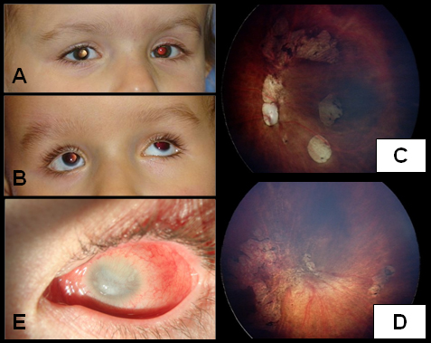
 Figure 1 of
Meire, Mol Vis 2011; 17:2072-2079.
Figure 1 of
Meire, Mol Vis 2011; 17:2072-2079.