Figure 2. TGF-β induces expression of
SPARC in human Tenon’s fibroblasts in vitro. Representative northern
blot analysis (A, B) and densitometry (C) for
SPARC expression in HTF cells without and with incubation with
activated TGF-β1 (1 ng/ml) or TGF-β2 (1 ng/ml) for three days. For
quantification, SPARC mRNA levels were measured
densitometrically, normalized to 28S methylene blue staining and
expressed as ×-fold to levels of untreated control cells (mean±SEM of 3
independent experiments; *p<0.05). Representative western blot
analysis (D, E) and densitometry (F) in HTF cells
for SPARC without and with incubation with activated TGF-β1 (1 ng/ml)
or TGF-β2 (1 ng/ml) for three days. For quantification, SPARC protein
levels were measured densitometrically, normalized to coomassie blue
staining and expressed as ×-fold to levels of untreated control cells
(mean±SEM of 3 independent experiments; *p<0.05).
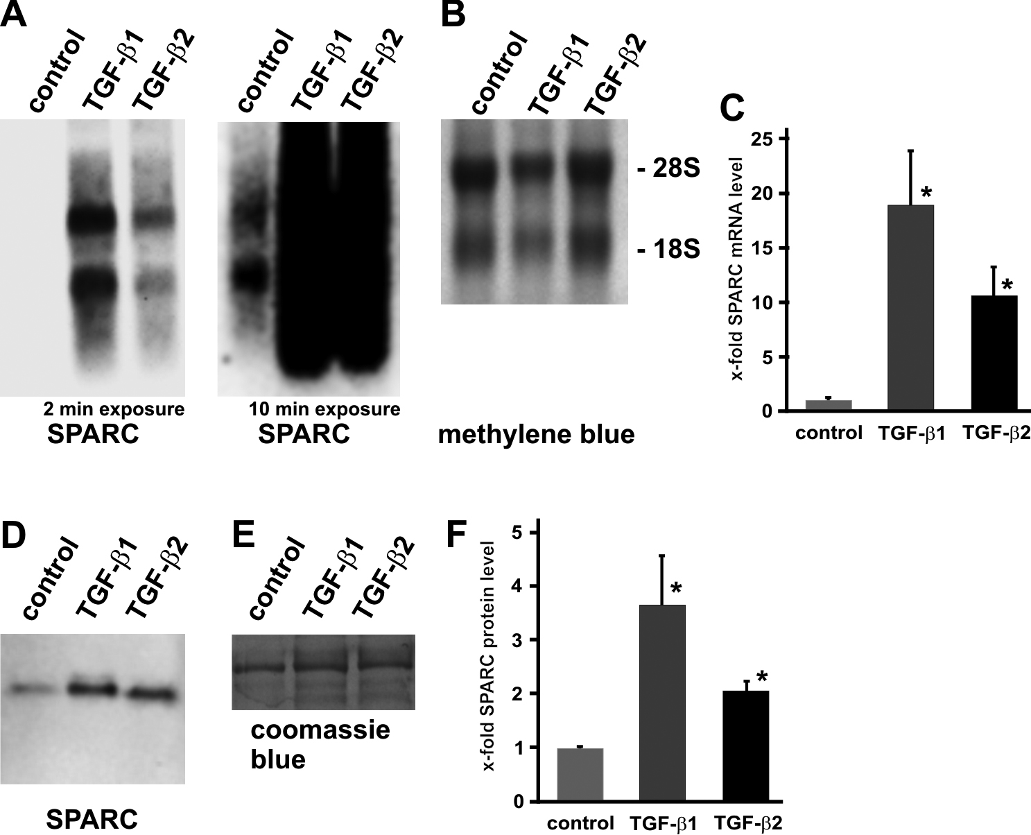
 Figure 2 of Fuchshofer, Mol Vis 2011; 17:177-185.