Figure 1. SPARC expression is increased in
human Tenon’s capsule scars. Immunohistochemistry for SPARC (green) in
scarred human Tenon’s capsule (C) shows a marked accumulation of
SPARC around and within vessel walls (arrows) and in areas of condensed
extracellular matrix (arrow heads), whereas only weak signals are
detected in extracellular matrix and vessel walls (arrows) of healthy
tissue (B). A: control immunostaining of a healthy
Tenon’s capsule. red: propidium iodide staining; original
magnification: 20×.
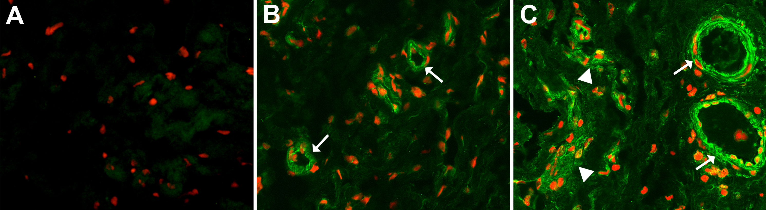
 Figure 1 of Fuchshofer, Mol Vis 2011; 17:177-185.